Anti-HDAC-1 antibody was prepared from whole rabbit serum produced by repeated immunizations with a synthetic peptide corresponding to a C-Terminal region near amino acids 450-482 of Human HDAC-1.
Anti-HDAC-1 Antibody has been tested for use in ELISA, immunohistochemistry, immunofluorescence, and western blot. Specific conditions for reactivity should be optimized by the end user. Specific nuclear staining is observed by IHC. Expect bands at 65 kD
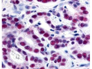
Immunohistochemistry of Rabbit Anti-HDAC-1 Antibody. Tissue: human prostate cancer tissue. Fixation: formalin fixed paraffin embedded. Antigen retrieval: not required. Primary antibody: HDAC-1 antibody at 1:500 for 1 h at RT. Secondary antibody: Peroxidase rabbit secondary antibody at 1:10,000 for 45 min at RT. Localization: HDAC-1 is nuclear. Staining: HDAC-1 precipitated purple with blue counterstain. Personal Communication, Alan Yen, LifeSpanBiosciences, Seattle, WA.
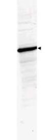
Western Blot of Rabbit Anti-HDAC-1 Antibody. Lane 1: 293 whole cell lysate (p/n W09-000-365). Load: 35 µg per lane. Primary antibody: HDAC-1 antibody at 1:3,500 for overnight at 4C. Secondary antibody: IRDye800(TM) rabbit secondary antibody at 1:10,000 for 45 min at RT. Block: 5% BLOTTO (p/n B501-0500) overnight at 4C. Predicted/Observed size: ~65 kDa corresponding to human HDAC1. Other band(s): none.
Immunohistochemistry of Rabbit Anti-HDAC-1 Antibody. Tissue: human lung tissue. Fixation: formalin fixed paraffin embedded. Antigen retrieval: not required. Primary antibody: HDAC-1 antibody at 10 µg/mL for 1 h at RT. Secondary antibody: Peroxidase rabbit secondary antibody at 1:10,000 for 45 min at RT. Localization: HDAC-1 is nuclear. Staining: HDAC-1 as brown color indicates presence of protein, blue color shows cell nuclei.
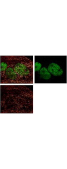
Immunofluorescence Microscopy of Rabbit Anti-HDAC-1 antibody. Fixation: 0.5% PFA. Antigen retrieval: not required. Primary antibody: HDAC-1 antibody at 10 µg/mL for 1 h at RT. Secondary antibody: rabbit secondary antibody at 1:10,000 for 45 min at RT. Localization: HDAC-1 is nuclear. Staining: HDAC-1 was used with Atto 425 (shown in red). Anti-Keratin monoclonal antibody was used with Dylight 488 (shown in green) to detect Keratin. Data was collected on a STED-CW TCS-SP5 Confocal system (Leica Microsystems).
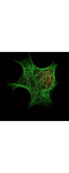
Immunofluorescence Microscopy of Rabbit anti-HDAC1 Antibody. Tissue: A431 cells. Fixation: methanol. Antigen retrieval: blocked with normal goat serum. Primary antibody: HDAC1 antibody at 4 µg/mL for 1 h at RT. Secondary antibody: rabbit secondary antibody at 0.2 µg/mL for 45 min at RT. Localization: HDAC1 is nuclear. Staining: HDAC1 as green fluorescent signal. A-tubulin monoclonal antibody detected with ATTO 425 (colored RED). 2-color STED image, Leica Microsystems.
Western Blot of Rabbit Anti-HDAC-1 Antibody. Lane 1: LNCaP prostate cancer cells. Load: 50 µg per lane. Primary antibody: HDAC-1 antibody at 1:1000 for overnight at 4C. Secondary antibody: IRDye800(TM) rabbit secondary antibody at 1:10,000 for 45 min at RT. Block: 5% BLOTTO overnight at 4C. Predicted/Observed size: 55kDa for HDAC-1.
* VAT and and shipping costs not included. Errors and price changes excepted