This affinity purified antibody was prepared from whole rabbit serum produced by repeated immunizations with a synthetic peptide corresponding to an N-Terminal region near amino acids 15-40 of human MCM2 protein.
Conjugation:
Unconjugated
Alternative Names:
rabbit anti-MCM2 antibody, MCM-2, BM28, CCNL1, CDCL1, Minichromosome maintenance complex component 2, Nuclear protein BM28, DNA replication licensing factor MCM2
0.02 M Potassium Phosphate, 0.15 M Sodium Chloride, pH 7.2
Form:
Liquid (sterile filtered)
Target:
Human
Antibody Type:
Primary Antibody
Application Dilute:
ELISA: 1:10,000 - 1:50,000, WB: 1:500 - 1:2,000
Application Notes:
This affinity purified antibody has been tested for use in ELISA and by western blot. Specific conditions for reactivity should be optimized by the end user. Expect a band approximately 100 kDa in size corresponding to phosphorylated or unphosphorylated
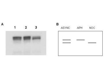
Western blot using Rocklands Affinity Purified anti-MCM2 antibody shows detection of both phosphorylated and unphosphorylated MCM2 present in nuclear extracts from elutriated human cells (MO59K/K562). The MCM2 protein is phosphorylated after initiation of DNA replication, therefore, the protein is unphosphorylated in early S phase, and gradually becomes phosphorylated throughout S phase. In G2/M, all MCM2 is phosphorylated. Panel A shows western blot results for lysates were prepared from asynchronous cells (lane 1), cells arrested in early S with aphidicolin (lane2), and cells arrested in mitosis with nocodazole (lane 3). Panel B shows a schematic diagram of bands representing phosphorylated and unphosphorylated MCM2 present in these preparations. Asynchronous cells contain a doublet of both forms. Aphidicolin treated cells contain only unphosphorylated MCM2 and nocodazole treatment results in only phosphorylated MCM2 detected in the lysate. The phosphorylated band migrates faster than the unphosphorylated form and is seen as the lower band. There is a clear switch from the unphosphorylated form in the center lane, to the phosphorylated form in the third lane, confirming recognition of both forms of MCM2 by this antibody. The primary antibody was diluted 1:400 for this experiment. Personal Communication, Jennifer Seiler, NIH, CCR, Bethesda, MD.
* VAT and and shipping costs not included. Errors and price changes excepted