0.02 M Potassium Phosphate, 0.15 M Sodium Chloride, pH 7.2
Form:
Liquid (sterile filtered)
Antibody Type:
Primary Antibody
Application Dilute:
ELISA: 1:1,000-1:2,500, Flow Cytometry: User Optimized, IHC: User Optimized, IF Microscopy: User Optimized, IP: 10 µg/mg protein sample, WB: 1:500-1:1,000
Application Notes:
Anti-Lysine Acetylated Antibody is suitable for use in ELISA, western blotting, immunofluorescence microscopy, and immunoprecipitation assays. Although not tested, this antibody is likely functional in RIA, flow cytometry, and immunohistochemistry.
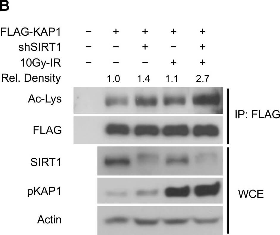
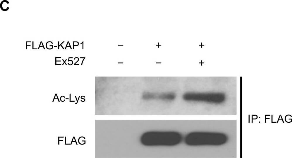
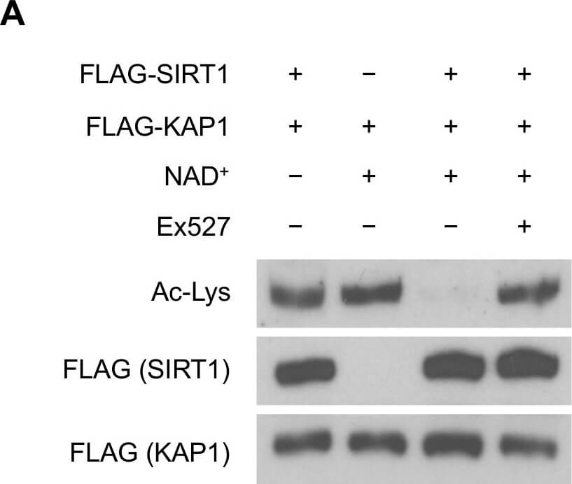
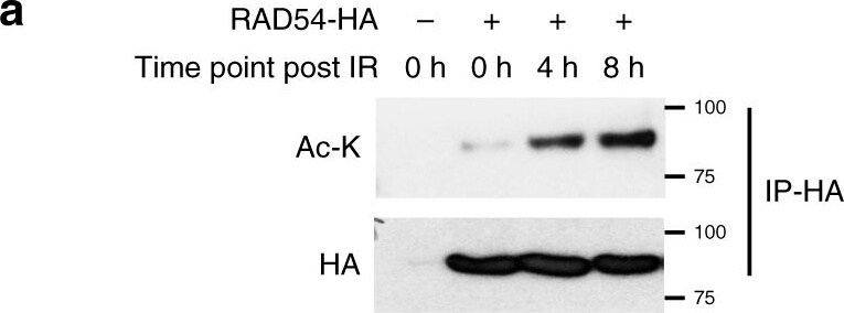
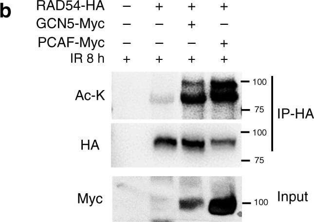
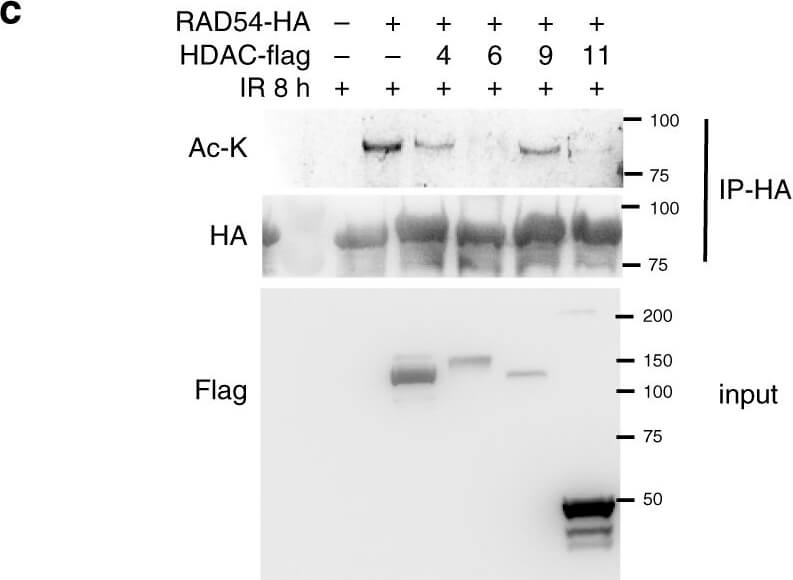
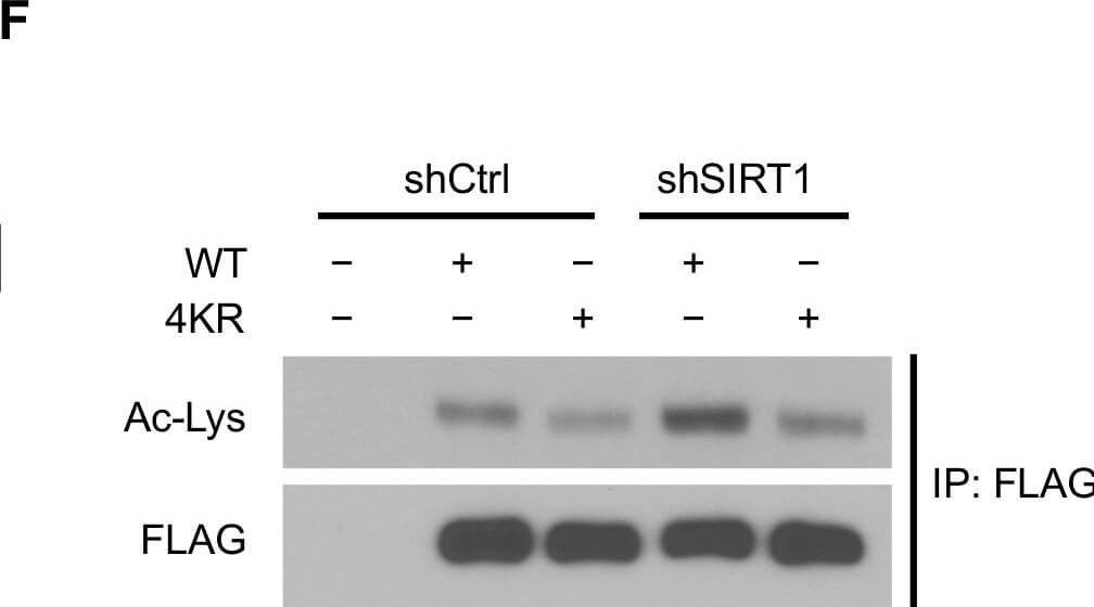
SIRT1 deacetylates KAP1 in vitro and in vivo.(A) SIRT1 deacetylates KAP1 in vitro. Exogenous FLAG-tagged KAP1 and SIRT1 were purified by anti-FLAG immunoprecipitation. Combination of purified proteins was incubated in deacetylation buffer supplemented with or without NAD+ cofactor. Ex527 (40µM) was added to block the deacetylase activity of SIRT1. (B) SIRT1 deacetylates KAP1 in vivo. FLAG-tagged KAP1 was transfected into control or SIRT1 depleted HEK293T cells. Cells were treated with or without IR 1 hour before harvest. Cell lysates were subjected to immunoprecipitation using anti-FLAG antibody, followed by western blot analysis to assess the total KAP1 acetylation level. Relative density of the overall acetyl lysine was quantified using ImageJ software. Acetylation level was normalized to corresponding FLAG band. (C) HEK293T cells transfected with FLAG-tagged KAP1 were treated with DMSO or Ex527 (20µM) for 6 hours before harvest. Cell lysates were then immunoprecipitated with FLAG-conjugated agarose beads, and the immunoprecipitates were blotted with anti-acetyl lysine antibody to determine the total acetylation level. (D and E) HEK293T cells were transfected with FLAG-tagged KAP1 and treated with DMSO or Ex527 (20µM) for 6 hours before harvest. Recombinant KAP1 was purified and sent for mass spectrometric analysis. Acetyl residues with >2-fold enhancement after inhibitor treatment were considered to be SIRT1 targeted sites. (F) Site-directed mutagenesis was applied to generate 4KR mutant. FLAG-tagged WT-KAP1 or 4KR mutant were transfected into control or SIRT1 depleted HEK293T cells. Total acetylation levels of recombinant WT-KAP1 and 4KR mutant were assessed by immunoblotting. Figure provided by CiteAb. Source: PLoS One, PMID: 25905708.
SIRT1 deacetylates KAP1 in vitro and in vivo.(A) SIRT1 deacetylates KAP1 in vitro. Exogenous FLAG-tagged KAP1 and SIRT1 were purified by anti-FLAG immunoprecipit
SIRT1 deacetylates KAP1 in vitro and in vivo.(A) SIRT1 deacetylates KAP1 in vitro. Exogenous FLAG-tagged KAP1 and SIRT1 were purified by anti-FLAG immunoprecipitation. Combination of purified proteins was incubated in deacetylation buffer supplemented with or without NAD+ cofactor. Ex527 (40µM) was added to block the deacetylase activity of SIRT1. (B) SIRT1 deacetylates KAP1 in vivo. FLAG-tagged KAP1 was transfected into control or SIRT1 depleted HEK293T cells. Cells were treated with or without IR 1 hour before harvest. Cell lysates were subjected to immunoprecipitation using anti-FLAG antibody, followed by western blot analysis to assess the total KAP1 acetylation level. Relative density of the overall acetyl lysine was quantified using ImageJ software. Acetylation level was normalized to corresponding FLAG band. (C) HEK293T cells transfected with FLAG-tagged KAP1 were treated with DMSO or Ex527 (20µM) for 6 hours before harvest. Cell lysates were then immunoprecipitated with FLAG-conjugated agarose beads, and the immunoprecipitates were blotted with anti-acetyl lysine antibody to determine the total acetylation level. (D and E) HEK293T cells were transfected with FLAG-tagged KAP1 and treated with DMSO or Ex527 (20µM) for 6 hours before harvest. Recombinant KAP1 was purified and sent for mass spectrometric analysis. Acetyl residues with >2-fold enhancement after inhibitor treatment were considered to be SIRT1 targeted sites. (F) Site-directed mutagenesis was applied to generate 4KR mutant. FLAG-tagged WT-KAP1 or 4KR mutant were transfected into control or SIRT1 depleted HEK293T cells. Total acetylation levels of recombinant WT-KAP1 and 4KR mutant were assessed by immunoblotting. Figure provided by CiteAb. Source: PLoS One, PMID: 25905708.
Rocklands Affinity Purified anti-Acetylated Lysine (AcK) antibody is shown to detect acetylated histone in TSA-treated mouse spleen cell lysate (Panel A), control (left lane) and TSA-treated mouse spleen cell lysate (right lane) in panel B, and in acetylated BSA loaded as indicated (panel C).
* VAT and and shipping costs not included. Errors and price changes excepted