
MyD88 Antibody, Rabbit, Polyclonal
Catalog Number:
ROC-600-401-955
- Images (10)
| Article Name: | MyD88 Antibody, Rabbit, Polyclonal |
| Biozol Catalog Number: | ROC-600-401-955 |
| Supplier Catalog Number: | 600-401-955 |
| Alternative Catalog Number: | ROC-600-401-955 |
| Manufacturer: | Rockland Immunochemicals |
| Host: | Rabbit |
| Category: | Antikörper |
| Application: | ELISA, IF, IHC, IP, WB |
| Species Reactivity: | Human, Mouse, Rat |
| Immunogen: | MYD88 Antibody was prepared from whole rabbit serum produced by repeated immunizations with a synthetic peptide corresponding to a region near the carboxy terminus of human MyD88 protein. The immunogen is located within the last 50 amino acids of MYD88. |
| Conjugation: | Unconjugated |
| Alternative Names: | Myeloid differentiation marker 88 antibody, Myeloid differentiation primary response gene 88 antibody, Myeloid differentiation primary response gene antibody, Myeloid differentiation primary response protein MyD88 antibody |
| Application Dilute: | ELISA: 1:5,000 - 1:20,000, IHC: 1:300 - 1:1000, IF Microscopy: 20 µg/mL, WB: 1:500 - 1:2,000 |
| Application Notes: | MYD88 Antibody has been tested for use in ELISA, immunofluorescence, immunoprecipitation, and western blot. This antibody is suitable for use in IHC and ICC. Expect a band approximately 33 kDa in size corresponding to MyD88 protein by western blotting in |
|
|
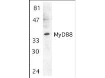
Western blot using Rocklands affinity purified anti-MyD88 antibody shows detection of MyD88 in Jurkat whole cell lysate. The membrane was probed with the primary antibody diluted to 1:500. Background Information: MyD88 (Myeloid differentiation primary response protein). The pro-inflammatory cytokine IL-1 induced cellular response requires IL-1 receptor complex including IL-1RI and IL-1RAcP. Recently, MyD88 was identified as an adapter molecule in the IL-1 signaling pathway (1). MyD88 associates with and recruits IRAK to the IL-1 receptor complex in response to IL-1 treatment and the dominant negative form of MyD88 attenuates IL-1R-mediated NF-kB activation. MyD88 is also employed as a regulator molecule by IL-18 receptor and human Toll receptor (2,3), both members of the Toll/IL-1R family of receptors. Targeted disruption of the MyD88 gene results in loss of cellular responses to IL-1 and IL-18, and MyD88-deficient mice lack responses to the bacterial product LPS that employs Toll-like receptors 2 and 4 (TLR2 and TLR4) as the signaling receptors. MyD88 is a general adapter protein for the Toll/IL-1R family of receptors and plays an important role in the inflammatory response induced by cytokines IL-1 and IL-18 and endotoxin. The MyD88 gene is expressed in many tissues. |
|
|
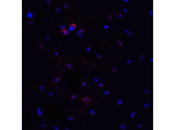
Immunofluorescence Validation of MyD88. Cell: Jurkat Cells. Fixation: 4% paraformaldehyde-fixed. Labeling: MyD88 at 20 µg/mL, followed by goat anti-rabbit IgG secondary antibody at 1:500 dilution (red) and DAPI staining (blue). |
|
|
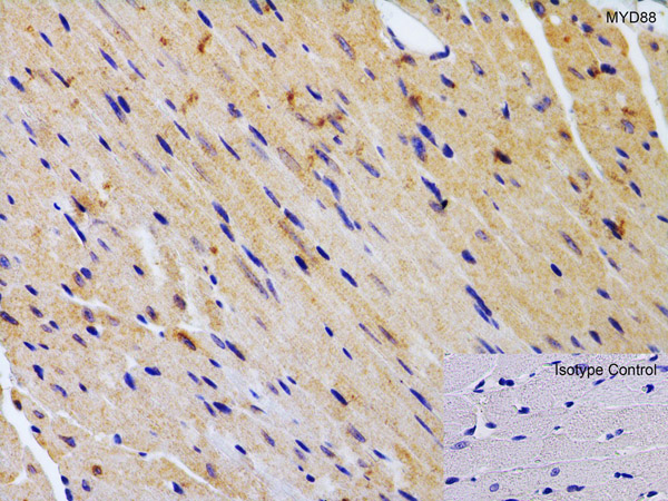
Immunohistochemistry Validation of MyD88. Tissue: Mouse Heart. Fixation: paraffin-embedded, formaldehyde and blocked with 10% serum for 1 h at RT. Antigen retrieval: heat mediation with a citrate buffer (pH6). Primary Antibody: anti-MYD88 antibody at 2 µg/ml overnight at 4C. Secondary: goat anti-rabbit IgG H&L (HRP) at 1:250. Counter stained with Hematoxylin. |
|
|
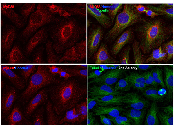
Immunofluorescence Validation of MyD88. Cell: HeLa Cells. Fixation: methanol-fixed. Labeling: MyD88 at 20 µg/mL, followed by goat anti-rabbit IgG secondary antibody at 1:1000 dilution (red) and Hoechst staining (blue). Alpha tubulin was stained with anti-alpha tubulin antibody following by goat anti-mouse IgG secondary antibody (green). Images were captured with confocal microscopy. |
|
|
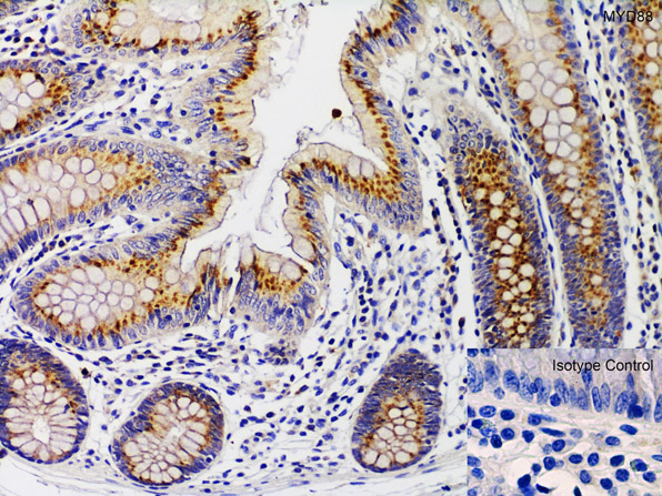
Immunohistochemistry Validation of MyD88. Tissue: Human Colon. Fixation: paraffin-embedded, formaldehyde and blocked with 10% serum for 1 h at RT. Antigen retrieval: heat mediation with a citrate buffer (pH6). Primary Antibody: anti-MYD88 antibody at 1 µg/ml overnight at 4C. Secondary: goat anti-rabbit IgG H&L (HRP) at 1:250. Counter stained with Hematoxylin. |
|
|
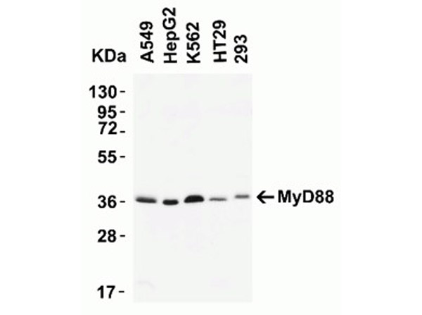
Western Blot Validation of MyD88. Load: 15 µg of human lysates per lane. Lane 1: A549, Lane 2: HepG2, Lane 3: K562, Lane 4: HT29, Lane 5: 293. Primary antibody: MyD88 at 2 µg/mL for 1 hr incubation at RT in 5% NFDM/TBST. Secondary: Goat anti-rabbit IgG HRP conjugate at 1:10000 dilution. Predicted band size: 35 kDa |
|
|
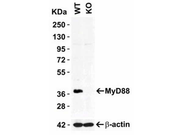
KO Western Blot Validation of MyD88.Load: 10 µg of HeLa WT cell lysate or MyD88 KO cell lysate. Primary antibody: MyD88 at 2 µg/mL and beta-actin 1 µg/mL for 1 hr incubation at RT in 5% NFDM/TBST.Secondary: Goat Anti-Rabbit IgG HRP conjugate at 1:10000 dilution. |
|
|
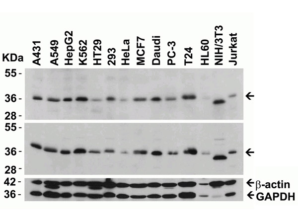
Western Blot Validation of MyD88. Load: 15 µg of lysates per lane. Lane 1: A431, Lane 2: A549, Lane 3: HepG2, Lane 4: K562, Lane 5: HT29, Lane 6: 293, Lane 7: HeLa, Lane 8: MCF7, Lane 9: Daudi, Lane 10: PC3, Lane 11: T24, Lane 12: HL60, Lane 13: 3T3/NIH, Lane 14: Jurkat. Primary antibody: MyD88 [competitor top] at 2 µg/mL, MyD88 [p/n 600-401-955 middle] at 2 µg/mL, beta-actin at 1 µg/mL, and GAPDH at 0.02 µg/mL for 1 hr incubation at RT in 5% NFDM/TBST. Secondary: Goat anti-rabbit IgG HRP conjugate at 1:10000 dilution. |
|
|
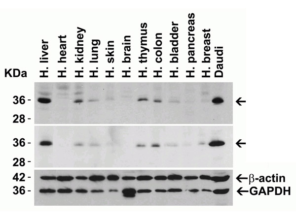
Western Blot Validation of MyD88. Load: 15 µg of human lysates per lane. Lane 1: liver, Lane 2: heart, Lane 3: kidney, Lane 4: lung, Lane 5: skin, Lane 6: brain, Lane 7: thymus, Lane 8: colon, Lane 9: bladder, La |
|
|
Product Guarantee and Expert Support
