This affinity purified antibody was prepared from whole rabbit serum produced by repeated immunizations with a synthetic peptide corresponding to an internal region of human AHA1 protein.
Conjugation:
Unconjugated
Alternative Names:
rabbit anti-AHA1 Antibody, Aha-1, Aha 1, Ahsa1 antibody, Activator of Hsp90 ATPase, Activator of 90 kDa heat shock protein ATPase homolog 1 antibody
0.02 M Potassium Phosphate, 0.15 M Sodium Chloride, pH 7.2
Form:
Liquid (sterile filtered)
Target:
Human
Antibody Type:
Primary Antibody
Application Dilute:
ELISA: 1:35,000 - 1:185,000, IHC: User Optimized, WB: 1 µg/mL
Application Notes:
This affinity purified antibody has been tested for use in ELISA and western blotting. Specific conditions for reactivity should be optimized by the end user. Expect a band approximately 38-40 kDa in size corresponding to AHA1 protein by western blotting
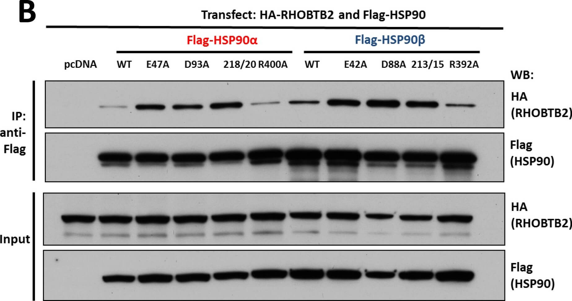
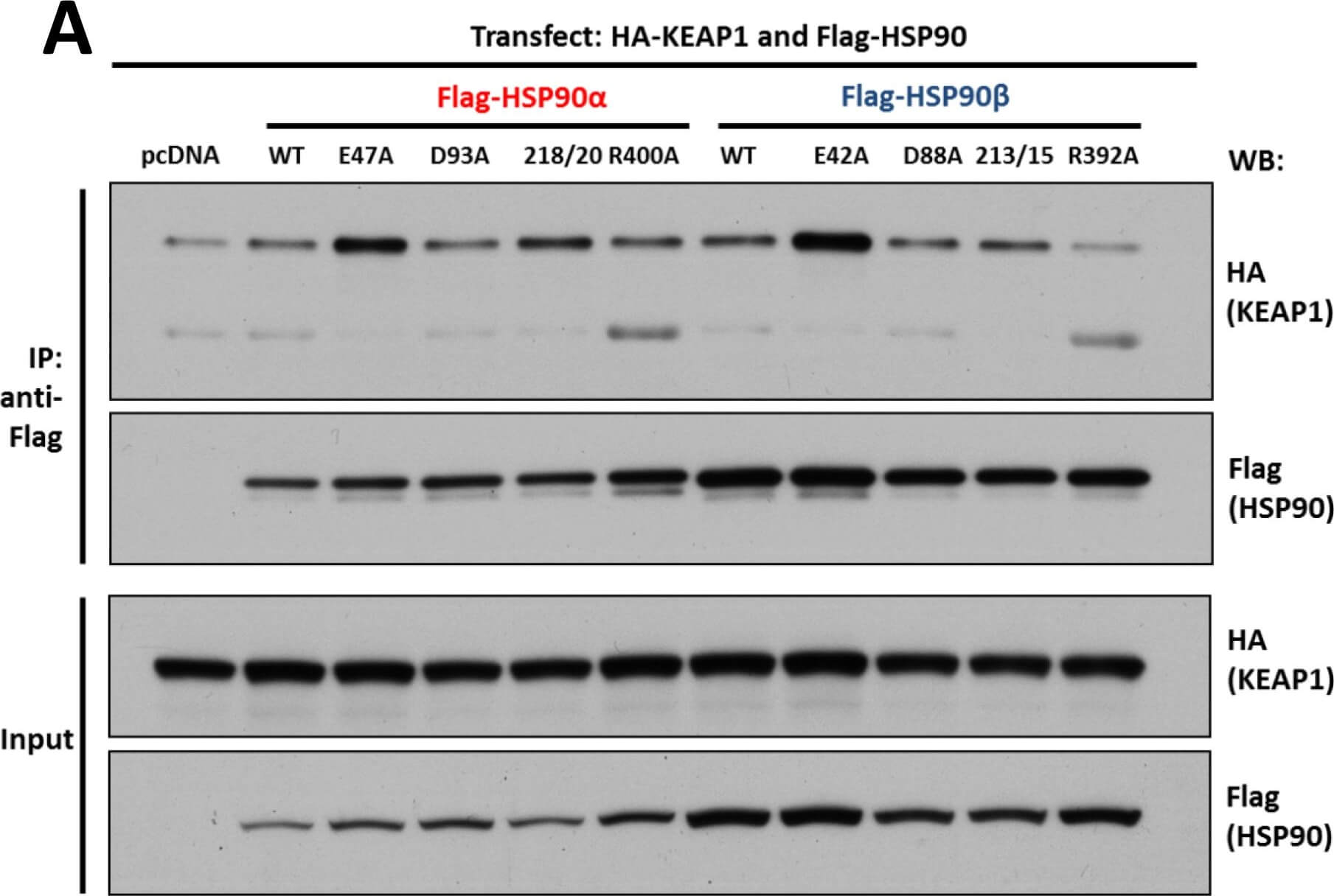
Interaction of KEAP1 and RHOBTB2 with HSP90 isoforms.(A) KEAP1interaction with HSP90 WT and mutants: HEK293 cells transfected with HA-KEAP1 and each FLAG-HSP90 construct were harvested, immunoprecipitated with anti-FLAG beads and western blotted for HA. Input lysates were normalized and run as controls. (B) RHOBTB2 interaction with HSP90 WT and mutants: HEK293 cells transfected with HA-RHOBTB2 and each FLAG-HSP90 construct were harvested, immunoprecipitated with anti-FLAG beads and western blotted for HA. Input lysates were normalized and run as controls. (C) Measurement of the relative interaction strength of KEAP1 and RHOBTB2 with each HSP90 isoform by LUMIER: HEK293 cells transfected with KEAP1 or RHOBTB2 and each HSP90 isoform were harvested, applied to a 96-well anti-FLAG plate and assayed for luciferase activity. The difference in relative interaction strength of HSP90alpha and HSP90beta for KEAP1 and RHOBTB2 (each approximately 3-fold) was statistically significant (p<0.05) (see Methods). Figure provided by CiteAb. Source: PLoS One, PMID: 26517842.
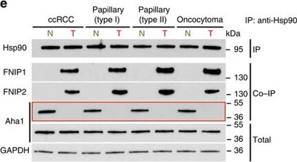
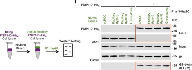
High levels of FNIPs make renal tumours sensitive to Hsp90 inhibitor GB.(a) Clear cell renal cell carcinoma (ccRCC), (b) Papillary type I, (c) Papillary type II, (d) Oncocytoma (Tumours, T) and adjacent normal tissues (Normal, N) were stained with haematoxylin and eosin (H&E). Proteins were also extracted from these tumours and adjacent normal tissues and incubated with indicated amounts of biotinylated GB followed by streptavidin agarose beads. Hsp90 was detected by immunoblotting. Expression of FNIP1 and FNIP2 in these samples was also detected by immunoblotting. (e
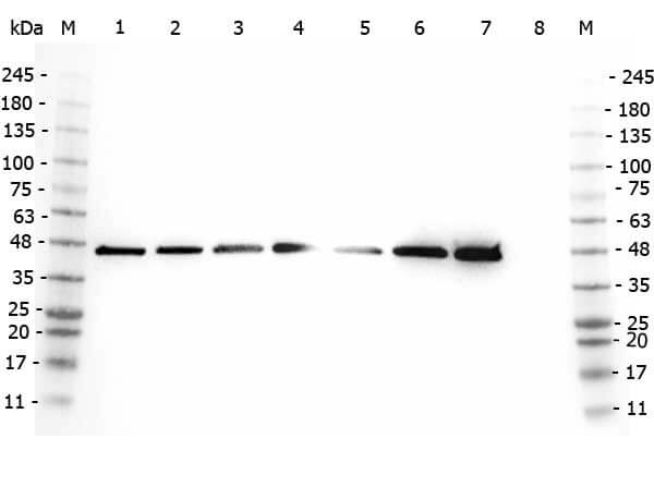
Western Blot of Rabbit anti-AHA1 antibody. Marker: Opal Pre-stained ladder (p/n MB-210-0500). Lane 1: HEK293 lysate (p/n W09-000-365). Lane 2: HeLa Lysate (p/n W09-000-364). Lane 3: MCF-7 Lysate (p/n W09-000-360). Lane 4: Jurkat Lysate (p/n W09-000-370). Lane 5: A431 Lysate (p/n W09-000-361). Lane 6: Raji Lysate (p/n W09-001-368). Lane 7: Ramos Lysate (p/n W09-000-GK4). Lane 8: NIH/3T3 Lysate (p/n W10-000-358). Load: 35 µg per lane. Primary antibody: AHA1 antibody at 1:2,000 for overnight at 4C. Secondary antibody: Peroxidase rabbit secondary antibody (p/n 611-103-122) at 1:30,000 for 60 min at RT. Blocking Buffer: 1% Casein-TTBS (p/n MB-082) for 30 min at RT. Predicted/Observed size: 38 kDa, 38 kDa for AHA1.
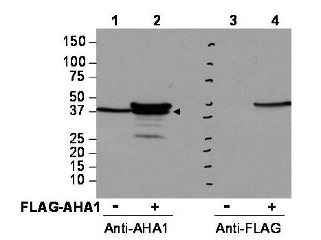
Western blot using Rocklands affinity purified anti-AHA1 antibody shows detection of AHA1 in Cos7 cells. For Lanes 2 and 4, Cos7 cells were transfected with pcDNA3-FLAG-AHA1. For Lanes 1 and 3, Cos7 cells were not transfected. Extracts (40 µg per lane) were electrophoresed and transferred to nitrocellulose. The membrane was probed with anti-AHA1 (lanes 1 and 2, 1:2,000 dilution) or anti-FLAG (lanes 3 and 4). The lower band seen in anti-AHA1 blotting (arrowhead) is endogenous AHA1. Personal Communication, Brad Scroggins, CCR-NCI, Bethesda, MD
Interaction of KEAP1 and RHOBTB2 with HSP90 isoforms.(A) KEAP1interaction with HSP90 WT and mutants: HEK293 cells transfected with HA-KEAP1 and each FLAG-HSP90 construct were harvested, immunoprecipitated with anti-FLAG beads and western blotted for HA. Input lysates were normalized and run as controls. (B) RHOBTB2 interaction with HSP90 WT and mutants: HEK293 cells transfected with HA-RHOBTB2 and each FLAG-HSP90 construct were harvested, immunoprecipitated with anti-FLAG beads and western blotted for HA. Input lysates were normalized and run as controls. (C) Measurement of the relative interaction strength of KEAP1 and RHOBTB2 with each HSP90 isoform by LUMIER: HEK293 cells transfected with KEAP1 or RHOBTB2 and each HSP90 isoform were harvested, applied to a 96-well anti-FLAG plate and assayed for luciferase activity. The difference in relative interaction strength of HSP90alpha and HSP90beta for KEAP1 and RHOBTB2 (each approximately 3-fold) was statistically significant (p<0.05) (see Methods). Figure provided by CiteAb. Source: PLoS One, PMID: 26517842.
* VAT and and shipping costs not included. Errors and price changes excepted