The immunogen is a mCherry mutant variant fusion protein of RFP corresponding to the full length amino acid sequence (234aa) derived from the mushroom polyp coral Discosoma.
Conjugation:
Unconjugated
Alternative Names:
rabbit anti-mCherry antibody, RFP, mCherry monomeric red fluorescent protein, Red Fluorescent Protein (RFP), rDsRed, Discosoma sp. Red Fluorescent Protein, mRFP1, mBanana, mHoneydew, mPlum, mOrange, mStrawberry, mTangerine
Clonality:
Polyclonal
Concentration:
1.17 mg/mL by UV absorbance at 280 nm
Buffer:
0.02 M Potassium Phosphate, 0.15 M Sodium Chloride, pH 7.2
Polyclonal anti-mCherry is designed to detect mCherry, RFP, and its variants. Anti-mCherry (Discosoma sp.) has been tested by ELISA and Western blot and is intended for use in immunological assays including ELISA, western blotting, immunofluorescence, an
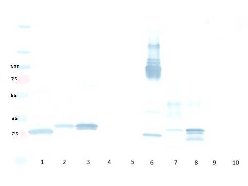
Western Blot of Rabbit anti-mCherry antibody. Lane 1: Rockland mCherry fusion protein (reduced). Lane 2: Control dsRed reduced. Lane 3: Control mCherry reduced. Lane 4: Control BFP reduced. Lane 5: Control eGFP reduced. Lane 6: Rockland mCherry fusion protein (non-reduced). Lane 7: Control dsRed non-reduced. Lane 8: Control mCherry non-reduced. Lane 9: Control BFP non-reduced. Lane 10: Control eGFP non-reduced. Total load ~200 nanograms per lane. Load: 200ng per lane. Primary Antibody: Anti-mCherry at 1:5000 for overnight at 4C. Secondary antibody: HRP rabbit secondary antibody at 1:10,000 and TMBM-100. Block: 5% BLOTTO overnight at 4C. Predicted/Observed size: 25.9 kDa, for mCherry and RFP, no reaction to BFP or GFP.
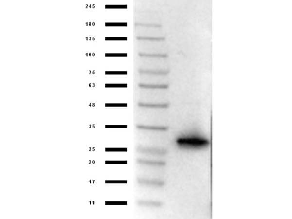
Western Blot of Rabbit Anti-mCherry Antibody. Lane 1: Opal Prestained Marker (p/n MB-210-0500). Lane 2: 50ng of RFP. Primary Antibody: rabbit anti-mCherry at 1µg/mL overnight at 4C. Secondary Antibody: goat anti-Rabbit peroxidase (p/n 611-103-122) at 1:70000 for 30mins at RT. Block: BlockOut Universal Buffer (p/n MB-073). Expect band ~30 kDa.
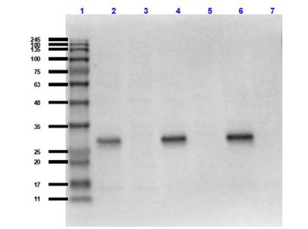
Western Blot of Rabbit Anti-mCherry Antibody MX Hu Ms Rt. Lane 1: Opal Prestained Molecular Weight Marker (p/n MB-210-0500). Lane 2: RFP (p/n 000-001-379)/HeLa WCL (p/n W09-000-364) [0.02µg/10µg]. Lane 3: HeLa WCL (p/n W09-000-364) [10µg]. Lane 4: RFP (p/n 000-001-379)/NIH/3T3 WCL (p/n W10-000-358) [0.02µg/10µg]. Lane 5: NIH/3T3 WCL (p/n W10-000-358) [10µg]. Lane 6: RFP (p/n 000-001-379)/PC-12 WCL (p/n W12-001-GL9) [0.02µg/10µg]. Lane 7: PC-12 WCL (p/n W12-001-GL9) [10µg]. Primary Antibody: Anti-mCherry at 1:1000 overnight at 2-8C. Secondary Antibody: Goat Anti-Rabbit IgG HRP (p/n 611-103-122) at 1:70,000 for 30mins at RT. Block: BlockOut Buffer (p/n MB-073). Predicted MW: ~27-30kDa.
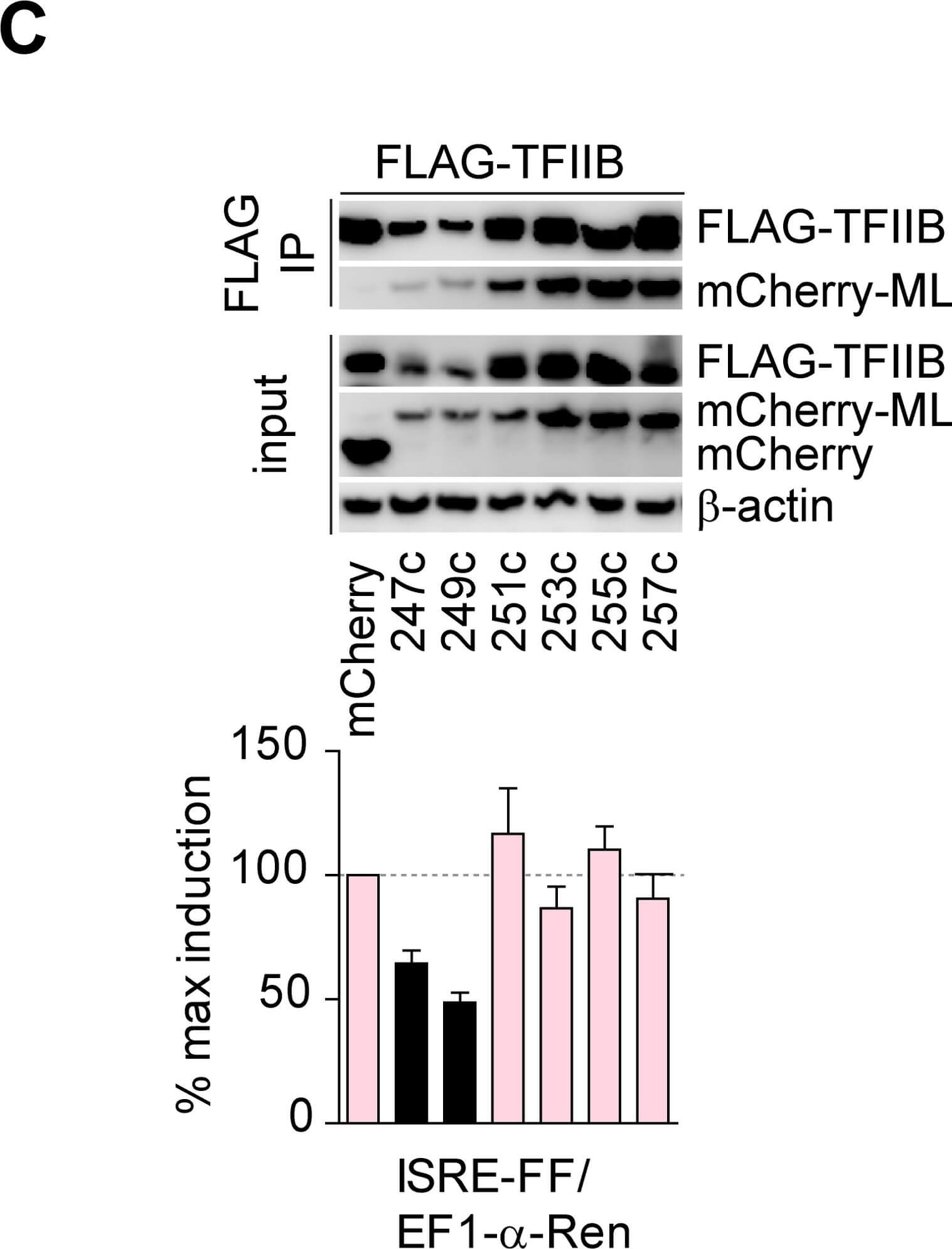
Mapping of a minimal ML sequence required for TFIIB inhibition.A) Schematic representation of ML fragments. B) Top panel: IP of FLAG-tagged TFIIB and GFP-fused ML fragments. Bottom panel: reporter assay in HEK293 cells, where Firefly luciferase under ISRE promoter was co-transfected with EF1-alpha-Renilla and GFP-ML fragments. C) Top-panel: IP of FLAG-tagged TFIIB and mCherry-fused ML fragments. Bottom panel: reporter assay in HEK293 cells, where Firefly luciferase under ISRE promoter was co-transfected with EF1-alpha-Renilla and mCherry-ML fragments. Western blots are representative of two experiments with similar results. Bar graphs show mean and SD from three technical replicates and are representative of two experiments with similar results. Figure provided by CiteAb. Source: PLoS Pathog, PMID: 29709033.
* VAT and and shipping costs not included. Errors and price changes excepted