PDL-1 Antibody was produced from whole rabbit serum prepared by repeated immunizations with a peptide corresponding to an internal region of human PDL-1.
0.02 M Potassium Phosphate, 0.15 M Sodium Chloride, pH 7.2
Form:
Liquid (sterile filtered)
Target:
Human
Antibody Type:
Primary Antibody
Application Dilute:
ELISA: 1:10,000 - 1:40,000, Flow Cytometry: 0.5 µg/ml, IHC: 2.5-5 µg/mL, IF Microscopy: User Optimized, WB: 0.5 - 1 µg/ml
Application Notes:
Rabbit Anti-PDL-1 antibody has been tested for use in ELISA, IHC, IF, FLOW, and western blotting. Expect a band at ~32 kDa in size corresponding to PDL-1 by western blotting in the appropriate cell lysate or extract. Raji cell lysate can be used as posit
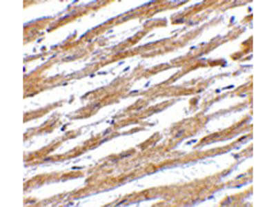
Immunohistochemistry of Rabbit anti-PDL-1 antibody. Tissue: human heart tissue. Fixation: formalin fixed paraffin embedded. Antigen retrieval: not required. Primary antibody: PDL1 antibody at 2.5 µg/mL for 1 h at RT. Secondary antibody: Peroxidase rabbit secondary antibody at 1:10,000 for 45 min at RT. Localization: PDL-1 is found in the cell membrane. Staining: PDL 1 as precipitated brown signal with hematoxylin purple nuclear counterstain.
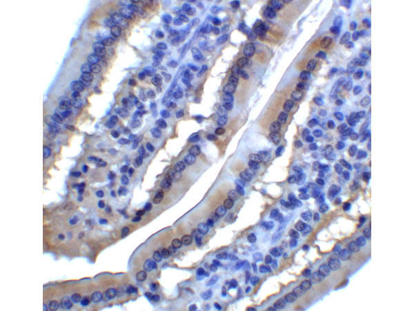
Immunohistochemistry of Rabbit anti-PDL-1 antibody. Tissue: Mouse Small Intestine. Fixation: formalin fixed paraffin embedded. Antigen retrieval: not required. Primary antibody: PDL1 antibody at 2 µg/mL for 1 h at RT. Secondary antibody: Biotinylated secondary antibody, HRP conjugated rabbit 1:200 for 30 min. Streptavidin-HRP for 30 min. Localization: PDL-1 is found in the cell membrane. Staining: PDL 1 as precipitated brown signal with hematoxylin purple nuclear counterstain.
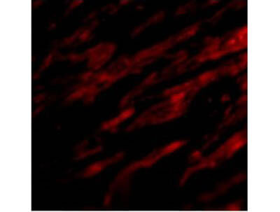
Immunofluorescence Microscopy of Rabbit anti-PDL-1 antibody. Tissue: human heart cells. Fixation: 0.5% PFA. Antigen retrieval: not required. Primary antibody: PDL 1 antibody at 20 µg/mL for 1 h at RT. Secondary antibody: Fluorescein rabbit secondary antibody at 1:10,000 for 45 min at RT. Localization: PDL-1 is found in the cell membrane. Staining: PDL1 as red signal.
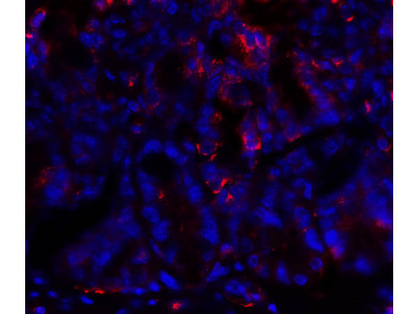
Immunofluorescence Microscopy of Rabbit anti-PDL-1 antibody. Tissue: Mouse Small Intestine. Fixation: 0.5% PFA. Antigen retrieval: not required. Primary antibody: PDL 1 antibody at 20 µg/mL for 1 h at RT. Secondary antibody: Fluorescein rabbit secondary antibody at 1:10,000 for 45 min at RT. Localization: PDL-1 is found in the cell membrane. Staining: PDL1 as red signal.
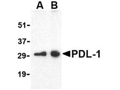
Western blot using Rocklands affinity purified anti-PDL-1. Lane A: Raji whole cell lysate at 0.5 µg/ml of anti-PDL-1 antibody. Lane B: Raji whole cell lysate at 1.0 µg/ml of anti-PDL-1 antibody. Expect: detection of a predominant band at ~31 kDa corresponding to PDL-1 (arrowhead).
* VAT and and shipping costs not included. Errors and price changes excepted