Anti-Caspase-12 antibody was prepared from whole rabbit serum produced by repeated immunizations with a synthetic peptide corresponding to amino acids within the large cleavage product of murine caspase-12. The immunogen is located within amino acids 170 - 220 of Caspase-12.
Anti-Caspase-12 Antibody (Large) has been tested for use in ELISA, Western Blotting, Immunohistochemistry and Immunofluorescence. Specific conditions for reactivity should be optimized by the end user. Expect a band at approximately 48 kDa in Western Blo
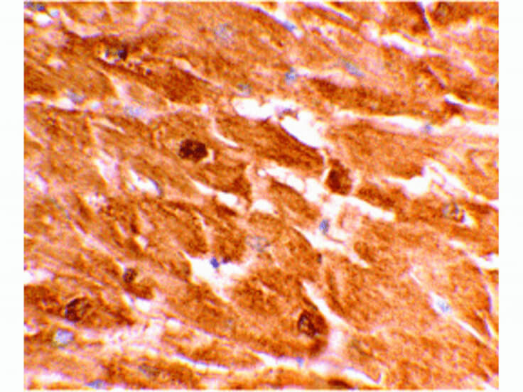
Immunohistochemistry of Capase-12 antibody. Tissue: human heart tissue. Fixation: formalin fixed paraffin embedded. Antigen retrieval: not required. Primary antibody: Capase-12 antibody at 2 µg/mL for 1 h at RT. Secondary antibody: Peroxidase rabbit secondary antibody at 1:10,000 for 45 min at RT. Localization: Capase-12 is endoplasmic reticular. Staining: Capase-12 is stained with hematoxylin purple nuclear counterstain.
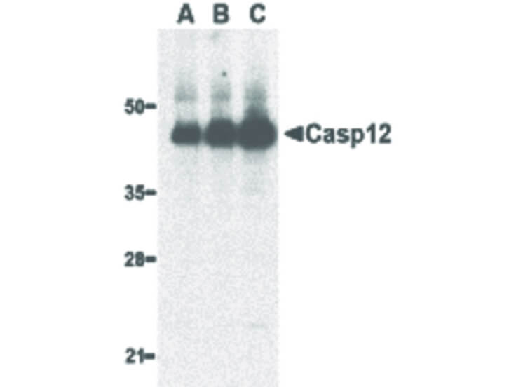
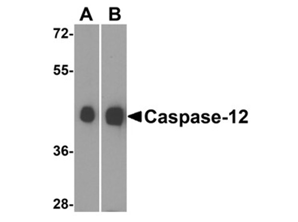
Western Blot of Caspase-12 antibody. Lane A: human heart lysate at 0.5 µg/ml. Lane B: human heart lysate at 1 µg/ml. Lane C: human heart lysate at 2 µg/ml. Load: 35 µg per lane. Primary antibody: Caspase-12 antibody at designated concentrations for overnight at 4C. Secondary antibody: Peroxidase rabbit secondary antibody at 1:10,000 for 45 min at RT. Block: 5% BLOTTO overnight at 4C. Predicted/Observed size: 48 kDa, 45 kDa for Caspase-12. Other band(s): Caspase-12 splice variants and isoforms.
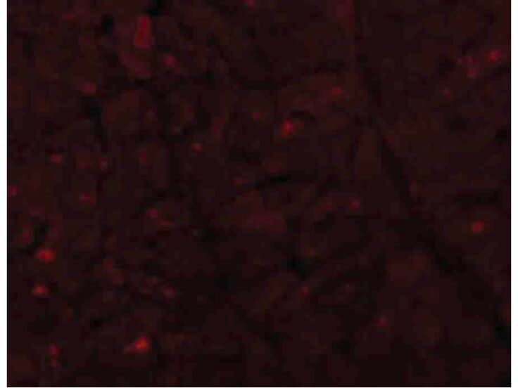
Immunofluorescence Microscopy of Caspase-12 antibody. Cell Type: human heart cells. Fixation: 0.5% PFA. Antigen retrieval: not required. Primary antibody: Caspase-12 antibody at 10 µg/mL for 1 h at RT. Secondary antibody: Fluorescein rabbit secondary antibody at 1:10,000 for 45 min at RT. Localization: Caspase-12 is endoplasmic reticular. Staining: Caspase-12 as red fluorescent signal.
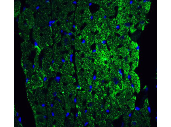
Immunofluorescence Validation of Caspase-12. Tissue: Mouse Heart. Fixation: 4% paraformaldehyde-fixed. Labeling: caspase-12 at 20 µg/mL, followed by goat anti-rabbit IgG secondary antibody at 1:500 dilution (green) and DAPI antibody (blue).
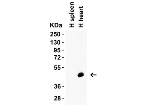
Western Blot Validation of Caspase 12. Load: 15 µg of human spleen or human heart lysate. Priamry antibody: Caspase-12 at 1 µg/mL for 1 h incubation at RT in 5% NFDM/TBST. Secondary: Goat Anti-Rabbit IgG HRP conjugate at 1:10000 dilution.
Western Blot Validation of Caspase 12. Load: 15 µg of lysate. Lane A: Human heart, Lane B: Mouse heart. Primary Antibody: Caspase-12 at 0.5 µg/mL for 1 h incubation at RT in 5% NFDM/TBST. Secondary: Goat Anti-Rabbit IgG HRP conjugate at 1:10000 dilution.
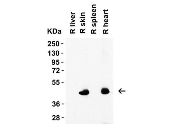
Western Blot Validation of Caspase 12. Load: 15 µg of rat lysates. Lane 1: rat liver, Lane 2: rat skin, Lane 3: rat spleen, Lane 4: rat heart. Primary antibody: Caspase-12 at 1 µg/mL for 1 h incubation at RT in 5% NFDM/TBST. Secondary: Goat Anti-Rabbit IgG HRP conjugate at 1:10000 dilution.
* VAT and and shipping costs not included. Errors and price changes excepted