Anti-Caspase-8 antibody was prepared from whole rabbit serum produced by repeated immunizations with a 16 amino acid synthetic peptide from near the C-terminus of human Caspase-8 isoform A. The immunogen is located within the last 50 amino acids of Caspase-8.
Anti-Caspase-8 Antibody has been tested for use in ELISA, Western Blotting, Immunocytochemistry, immunohistochemistry, and Immunofluorescence. Specific conditions for reactivity should be optimized by the end user. Expect a band at approximately 55 kDa i
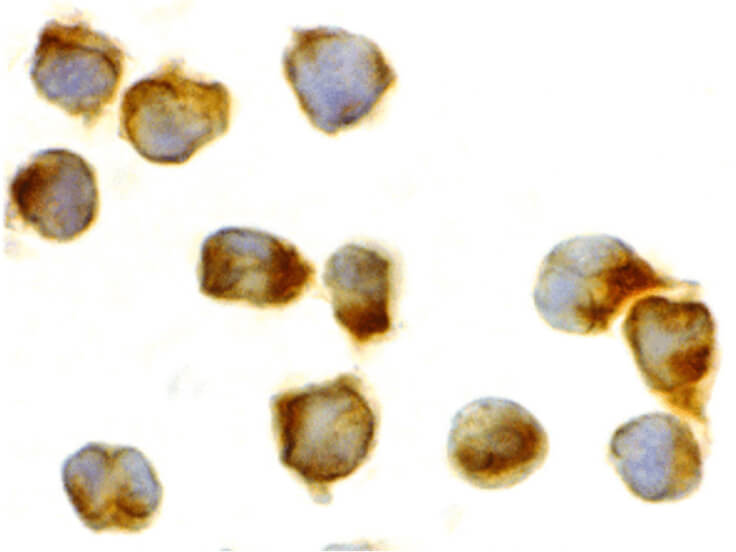
Immunocytochemistry of Caspase-8 antibody. Cell Type: Jurkat cells. Fixation: formalin fixed paraffin embedded. Antigen retrieval: not required. Primary antibody: Caspase-8 antibody at 2 µg/mL for 1 h at RT. Secondary antibody: Peroxidase rabbit secondary antibody at 1:10,000 for 45 min at RT. Localization: Caspase-8 is cytoplasmic. Staining: Caspase-8 as is stained brown with hematoxylin purple nuclear counterstain.
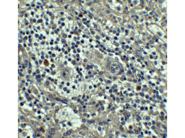
Immunohistochemistry of Caspase-8. Tissue: human spleen tissue. Primary antibody: Caspase-8 antibody at 5 µg/mL.
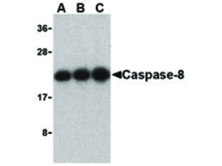
Western Blot of Caspase-8 antibody in Jurkat cell lysate. Lane A: Caspase-8 antibody at 0.5 µg/mL. Lane B: Caspase-8 antibody at 1 µg/mL. Lane C: Caspase-8 antibody at 2 µg/mL Load: 35 µg per lane. Primary antibody: Caspase-8 antibody at designated concentrations for overnight at 4C. Secondary antibody: Peroxidase rabbit secondary antibody at 1:10,000 for 45 min at RT. Block: 5% BLOTTO overnight at 4C. Predicted/Observed size: 55 kDa, 21 kDa for Caspase-8. Other band(s): Caspase-8 splice variants and isoforms.
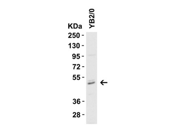
Western Blot Validation of Caspase 8. Load: 15 µg of rat YB2/0 cell lysate. Primary antibody: Caspase 8 at 1 µg/mL for 1 h incubation at RT in 5% NFDM/TBST. Secondary: Goat Anti-Rabbit IgG HRP conjugate at 1:10000 dilution.
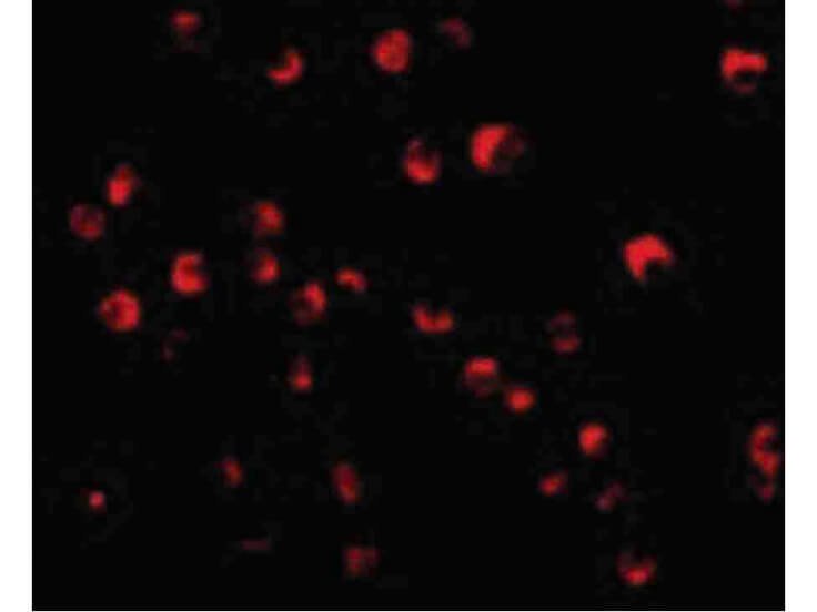
Immunofluorescence Microscopy of Caspase-8 antibody. Cell Type: Jurkat cells. Fixation: 0.5% PFA. Antigen retrieval: not required. Primary antibody: Caspase-8 antibody at 20 µg/mL for 1 h at RT. Secondary antibody: Fluorescein rabbit secondary antibody at 1:10,000 for 45 min at RT. Localization: Caspase-8 is cytoplasmic. Staining: Caspase-8 as red fluorescent signal.
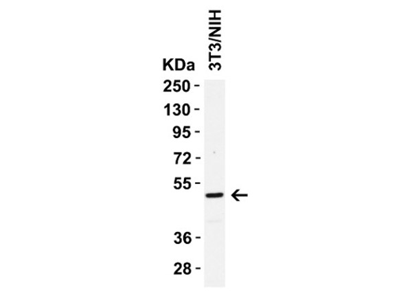
Western Blot Validation of Caspase 8.Load: 15 µg of 3T3/NIH cell lysate. Primary antibody: Caspase-8 at 1 µg/mL for 1 h incubation at RT in 5% NFDM/TBST. Secondary: Goat Anti-Rabbit IgG HRP conjugate at 1:10000 dilution.
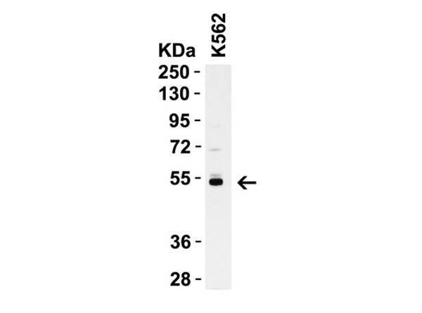
Western Blot Validation of Caspase 8. Load: 15 µg of K562 cell lysate. Primary antibody: Caspase 8 at 1 µg/mL for 1 h incubation at RT in 5% NFDM/TBST. Secondary: Goat Anti-Rabbit IgG HRP conjugate at 1:10000 dilution.
* VAT and and shipping costs not included. Errors and price changes excepted