This affinity purified antibody was prepared from whole rabbit serum produced by repeated immunizations with a phosphorylated synthetic peptide corresponding to the region of amino acids containing serine 206 of human SMAD1 protein.
Conjugation:
Unconjugated
Alternative Names:
rabbit anti-SMAD1 pS206 antibody, SMAD-1, SMAD 1, mothers against decapentaplegic homolog 1 antibody, MAD homolog 1, Mothers against DPP homolog 1, SMAD family member 1, MADH1, MADH 1, Transforming growth factor-beta-signaling protein 1, BSP-1, JV4-1
0.02 M Potassium Phosphate, 0.15 M Sodium Chloride, pH 7.2
Form:
Liquid (sterile filtered)
Target:
Human
Antibody Type:
Primary Antibody
Application Dilute:
ELISA: 1:5,000 - 1:25,000, WB: 1:500 - 1:2,000
Application Notes:
This affinity purified antibody has been tested for use in ELISA and by western blot. Specific conditions for reactivity should be optimized by the end user. Expect a band approximately 52 kDa in size corresponding to phosphorylated SMAD1 protein by west
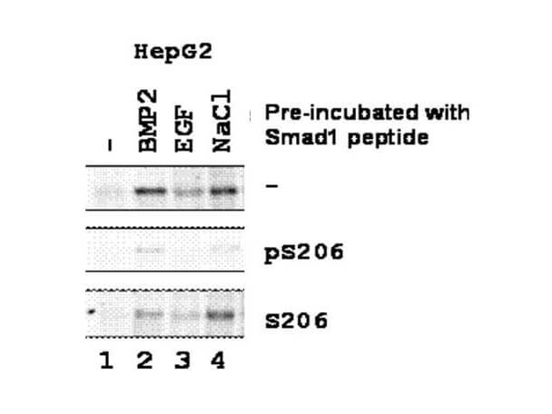
Western blot using Rocklands Affinity Purified anti-SMAD1 pS206 antibody shows detection of endogenous phosphorylated SMAD1 in whole cell lysates from human hepatoma (HEPG2, lanes 1-4) derived cell lines treated with PBS, BMP2 (5 ng/mL), EGF (1 ng/mL), or NaCl for 1 h at 37C before harvest. Each lane contains approximately 15 µg of lysate. Primary antibody was used at a 1:500 dilution in 1% BLOTTO (p/n B501-0500) and reacted for 1 hour at room temperature. Primary antibody was pre-incubated before reacting with blot as follows: top row - with PBS, middle row - with the immunizing phosphorylated peptide and bottom row - with control or non-phosphorylated peptide. The membrane was washed and reacted with a 1:3,000 dilution HRP-conjugated a-Rabbit IgG (p/n 611-103-122) for 1 hour at room temperature. Detection was by ECL. Personal communication, Xin-Hua Feng, Baylor College of Medicine, Houston, TX.
* VAT and and shipping costs not included. Errors and price changes excepted