Anti-GLS2 antibody was prepared from whole rabbit serum produced by repeated immunizations with a 18 amino acid synthetic peptide near the internal region terminus of human GLS2.
Anti-GLS2 Antibody has been tested for use in ELISA, Western Blotting, Immunohistochemistry and Immunofluorescence. Specific conditions for reactivity should be optimized by the end user. Expect a band at approximately 66 kDa in Western Blots of specific
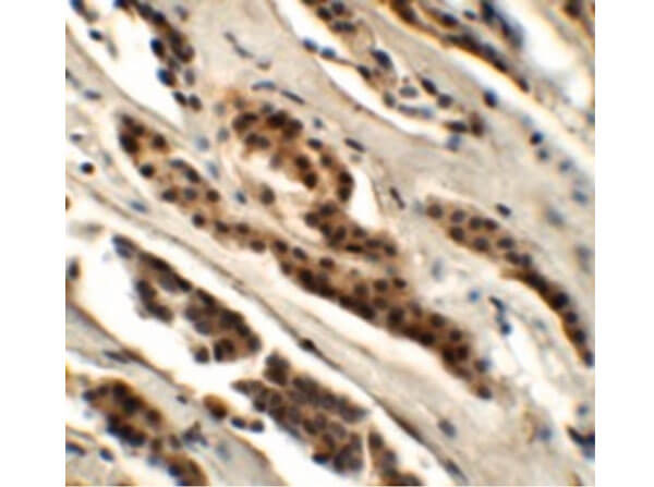
Immunohistochemistry of Rabbit anti-GLS2 antibody. Tissue: human kidney tissue. Primary antibody: GLS2 antibody at 5.0 µg/mL. Secondary antibody: Rabbit secondary antibody at 1:5,000 Localization: GLS2 is located in the mitochondrion. Staining: GLS2 as precipitated purple signal.
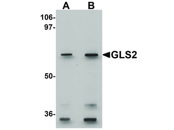
Western Blot of Rabbit anti-GLS2 antibody. Lane A: rat kidney tissue lysate at 0.5 µg/mL. Lane B: rat kidney tissue lysate at 1µg/mL. Primary antibody: GLS2 antibody overnight at 4C. Secondary antibody: anti-Rabbit HRP secondary antibody. Block: 5% BLOTTO. Predicted/Observed size: 66 kDa for GLS2 antibody.
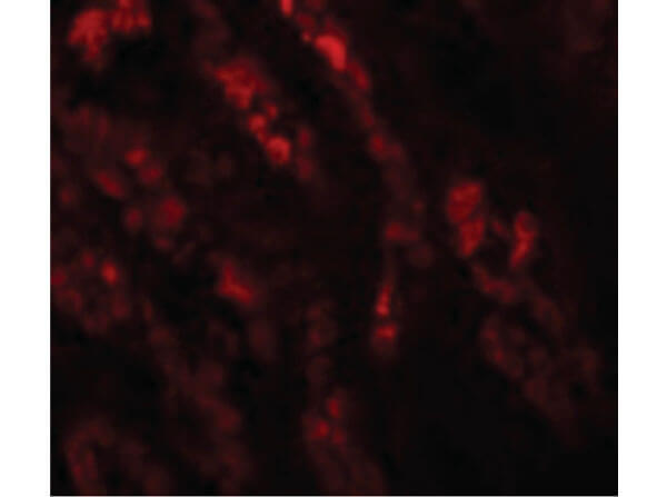
Immunofluorescence Microscopy of Rabbit anti-GLS2 antibody. Tissue: Human Kidney cells. Primary antibody: GLS2 antibody at 20 µg/mL. Secondary antibody: Fluorescein rabbit secondary antibody at 1:20,000. Localization: GLS2 is located in the mitochondrion. Staining: GLS2 as red fluorescent signal.
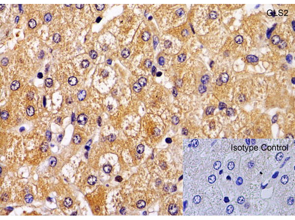
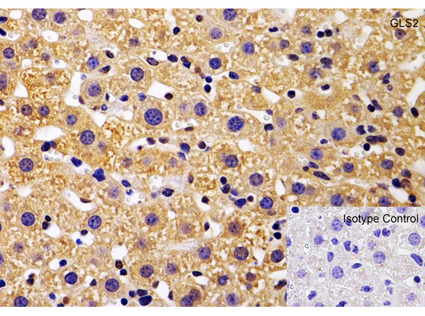
Immunohistochemistry Validation of GLS2.Tissue: Human Liver. Fixation: paraffin-embedded, formaldehyde and blocked with 10% serum for 1 h at RT. Antigen retrieval: heat mediation with a citrate buffer (pH6). Primary Antibody: anti-GLS2 antibody at 1 µg/ml overnight at 4C. Secondary: goat anti-rabbit IgG H&L (HRP) at 1:250. Counter stained with Hematoxylin.
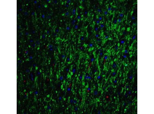
Immunofluorescence Validation of GLS2. Tissue: Mouse Brain. Fixation: 4% paraformaldehyde-fixed. Primary Antibody: GLS2 at 20 µg/mL. Secondary: goat anti-rabbit IgG antibody at 1:500 dilution (green) and DAPI antibody (blue).
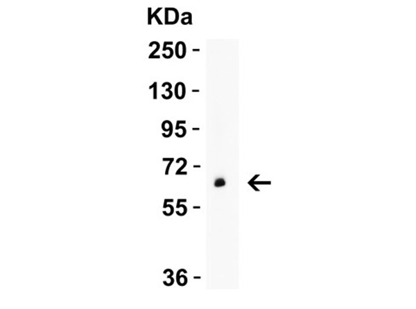
Western Blot Validation of GLS2.Load: 10 µg of human pancreas lysate. Primary Antibody: GLS2 at 2 µg/mL for 1 h incubation at RT in 5% NFDM/TBST. Secondary: Goat Anti-Rabbit IgG HRP conjugate at 1:10000 dilution.
Immunohistochemistry Validation of GLS2. Tissue: Mouse Liver. Fixation: paraffin-embedded, formaldehyde and blocked with 10% serum for 1 h at RT. Antigen retrieval: heat mediation with a citrate buffer (pH6). Primary Antibody: anti-GLS2 antibody at 1 µg/ml overnight at 4C. Secondary: goat anti-rabbit IgG H&L (HRP) at 1:250. Counter stained with Hematoxylin.
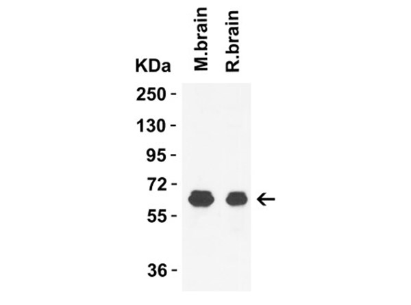
Western Blot Validation of GLS2. Load: 15 µg of mouse brain lysate (lane 1) or rat brain lysate (lane 2). Primary Antibody: GLS2 at 1 µg/mL for 1 h incubation at RT in 5% NFDM/TBST. Secondary: Goat Anti-Rabbit IgG HRP conjugate at 1:10000 dilution.
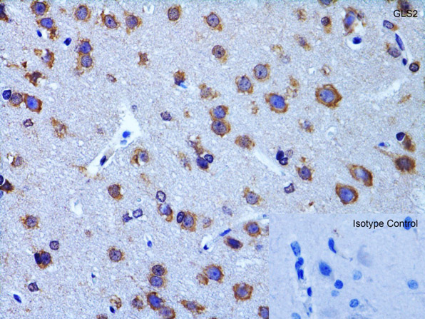
Immunohistochemistry Validation of GLS2. Tissue: Rat Brain. Fixation: paraffin-embedded, formaldehyde and blocked with 10% serum for 1 h at RT. Antigen retrieval: heat mediation with a citrate buffer (pH6).Primary Antibody: anti-GLS2 antibody at 2 µg/ml overnight at 4C. Secondary: goat anti-rabbit IgG H&L (HRP) at 1:250. Counter stained with Hematoxylin.
* VAT and and shipping costs not included. Errors and price changes excepted