Anti-XAF-1 antibody was prepared from whole rabbit serum produced by repeated immunizations with a synthetic peptide corresponding to amino acids at the C-terminus of human XAF-1.
Anti-XAF-1 Antibody has been tested for use in ELISA, Western Blotting, Immunohistochemistry and Immunofluorescence. Specific conditions for reactivity should be optimized by the end user. Expect a band at approximately 35 kDa in Western Blots of specifi
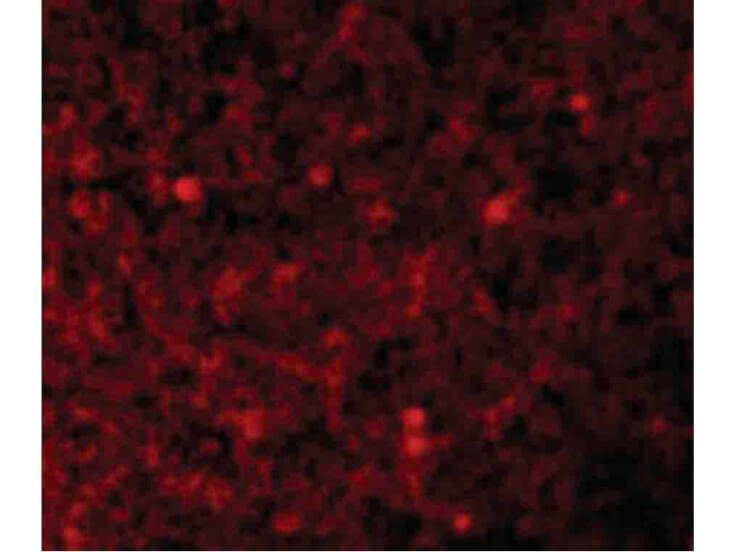
Immunofluorescence Microscopy of XAF-1 antibody. Tissue: human spleen cells. Fixation: 0.5% PFA. Antigen retrieval: not required. Primary antibody: XAF-1 antibody at 10 µg/mL for 1 h at RT. Secondary antibody: Fluorescein rabbit secondary antibody at 1:10,000 for 45 min at RT. Localization: XAF-1 is located in the cytoplasm, cytoplasmic vesicle, cytoskeleton, endosome, golgi apparatus, and cell membrane. Staining: XAF-1 as red fluorescent signal.
* VAT and and shipping costs not included. Errors and price changes excepted