ACE2 Antibody was produced from whole rabbit serum prepared by repeated immunizations with a synthetic peptide corresponding to amino acids near the C-terminus of human ACE2.
Conjugation:
Unconjugated
Alternative Names:
ACE2 Antibody, ACEH, Angiotensin-converting enzyme 2, ACE-related carboxypeptidase, ACEH
0.02 M Potassium Phosphate, 0.15 M Sodium Chloride, pH 7.2
Form:
Liquid (sterile filtered)
Target:
Human
Antibody Type:
Primary Antibody
Application Dilute:
ELISA: User Optimized, IHC: 2µg/mL, IF Microscopy: 10µg/mL, WB: 0.5-2µg/mL
Application Notes:
Anti-ACE2 Antibody is tested for use in E, WB, IF, and IHC. Expect a band approximately ~92.4 kDa on specific lysates. Western Blot validated in human, mouse and rat samples. Immunohistochemistry and Immunofluorescence validated in human samples. Specifi
Immunofluorescene of Rabbit Anti-ACE2 Antibody. Tissue: Human Lung Tissue. Fixative: 4% PFA. Primary Antibody: Anti-ACE2 at 20µg/mL. Secondary Antibody: Goat anti-rabbit IgG secondary antibody at 1:500 dilution (green) and DAPI counterstain (blue).
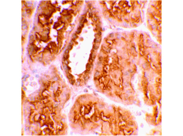
Immunohistochemistry of Anti-ACE2 Antibody. Tissue: Human Kidney Tissue. Fixation: formaldahyde and blocked with 10% serum for 1hr at RT. Antigen retrieval: heat mediation with Citrate Buffer pH6. Primary antibody: ACE2 antibody at 2µg/mL for overnight at 2-8C. Secondary antibody: Goat Anti-rabbit HRP secondary antibody 1:250 for 45 min at RT. Counterstain: hematoxylin.
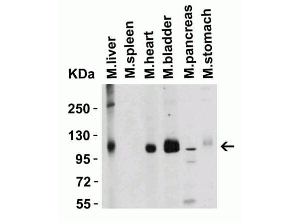
Western Blot of ACE2 Antibody. Lane 1: Mouse Liver Lysate. Lane 2: Mouse Spleen Lysate. Lane 3: Mouse Heart Lysate. Lane 4: Mouse Bladder Lysate. Lane 5: Mouse Pancreas Lysate. Lane 6: Mouse Stomach Lysate. Loading: 15 µg of lysates per lane. Primary Antibody: Anti-ACE2 at 2µg/mL for 1hr at RT in 5% BLOTTO/TBST. Secondary Antibody: Goat anti-rabbit IgG HRP conjugate at 1:10000 dilution. Predicted MW: ~93kDa. Observed MW: ~125kDa.
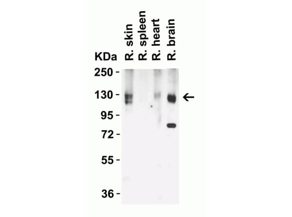
Western Blot of Rb Anti-ACE2 Antibody. Lane 1: Rat Skin Lysate. Lane 2: Rat Spleen Lysate. Lane 3: Rat Heart Lysate. Lane 4: Rat Brain Lysate. Loading: 15 µg of lysates per lane. Primary Antibody: Anti-ACE2 at 2µg/mL for 1hr at RT in 5% BLOTTO/TBST. Secondary: Goat anti-rabbit IgG HRP conjugate at 1:10000 dilution. Predicted MW: ~93kDa. Observed MW: ~130kDa.
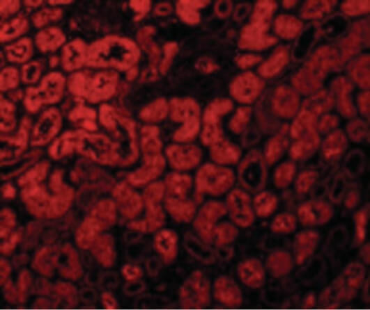
Immunofluorescence Microscopy of Rabbit Anti-ACE2 antibody. Tissue: Human Kidney Tissue. Fixation: 4% PFA. Primary antibody: ACE2 antibody at 10 µg/mL for overnight at 2-8C. Secondary antibody: Goat Anti-Rabbit IgG Conjugated 1:500 for 1hr at RT.
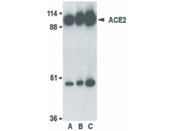
Western Blot of Rabbit Anti-ACE2 Antibody. Load: Human Kidney Lysate. Primary Antibody at - Lane A: 0.5µg/mL, Lane B: 1.0µg/mL, Lane C: 2.0 µg/mL. Expect: ~92kDa.
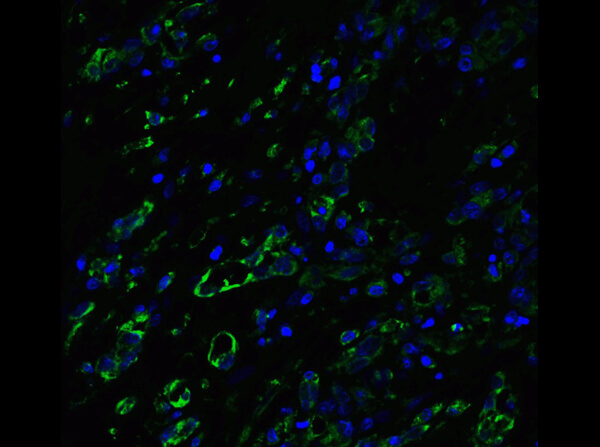
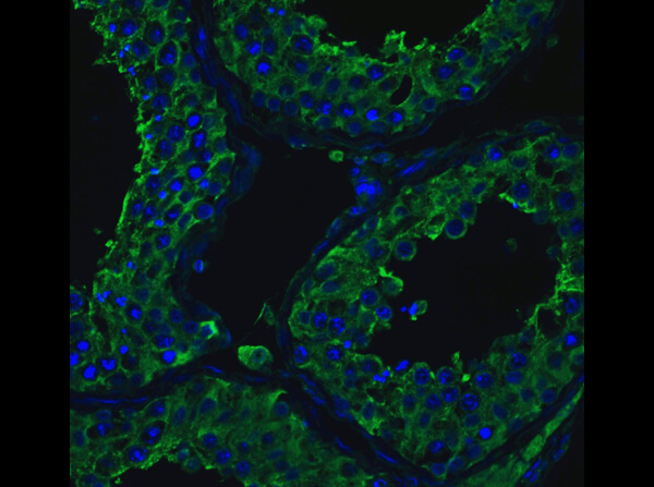
Immunofluorescence of Rabbit Anti-ACE2 Antibody. Tissue: Human Testis. Fixative: 4% PFA. Primary Antibody: Anti-ACE2 at 20µg/mL. Secondary Antibody: Goat anti-rabbit IgG secondary antibody at 1:500 dilution (green) and DAPI counterstain (blue).
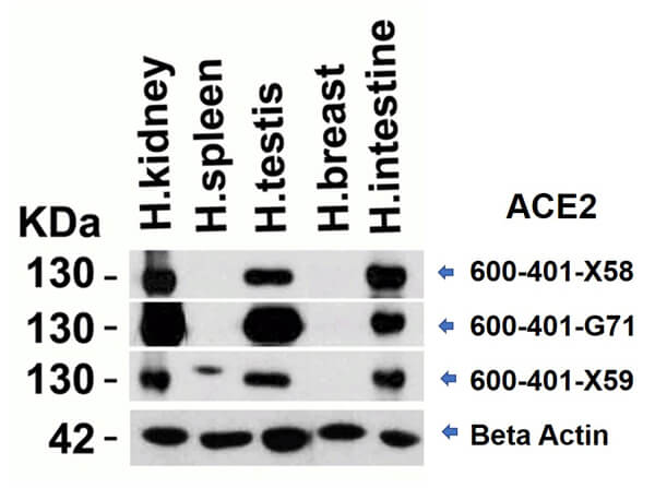
Western Blot of different Rabbit anti-ACE2 antibodies. Lane 1: Human Kidney Lysate. Lane 2: Human Spleen Lysate. Lane 3: Human Testis Lysate. Lane 4: Human Breast Lysate. Lane 5: Human Intestine Lysate. Load: 15 µg per lane. Primary antibody: ACE2 antibody (600-401-X58, 600-401-G71, 600-401-X59) at 2µg/mL and Beta Actin at 1µg/mL for 1hr at RT. Secondary antibody: Goat anti-Rabbit secondary HRP antibody. Block: 5% BLOTTO/TBST. Predicted MW: ~93kDa. Observed: ~130kDa.
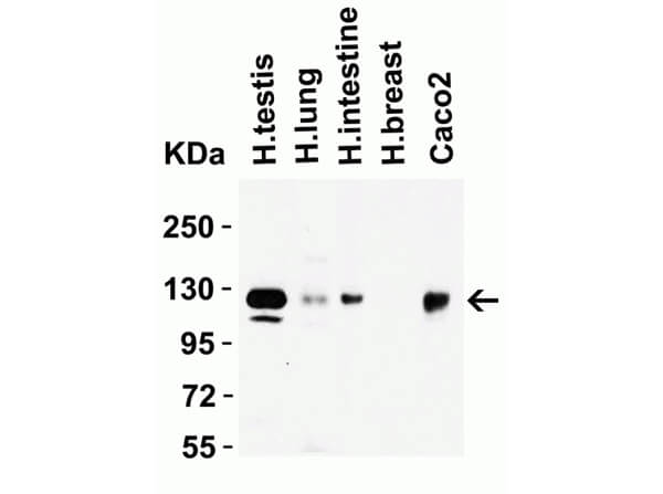
Western Blot of Rabbit Anti-ACE2 Antibody. Lane 1: Human Testis Lysate. Lane 2: Human Lung Lysate. Lane 3: Human Intestine Lysate. Lane 4: Human Breast Lysate. Lane 5: Caco2 Lysate. Loading: 15 µg of lysates per lane. Primary Antibody: Anti-ACE2 at 2µg/mL for 1hr at RT in 5% BLOTTO/TBST. Secondary Antibody: Goat anti-rabbit IgG HRP conjugate at 1:10000 dilution. Predicted MW: ~93kDa. Observed MW: ~130kDa.
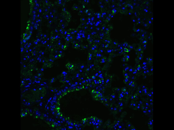
Immunofluorescence of Rb Anti-ACE2 Antibody. Tissue: Mouse Lung Tissue. Fixative: 4% PFA. Primary Antibody: Anti-ACE2 at 20µg/mL. Secondary Antibody: Goat anti-rabbit IgG secondary antibody at 1:500 dilution (green) and DAPI counterstain (blue).
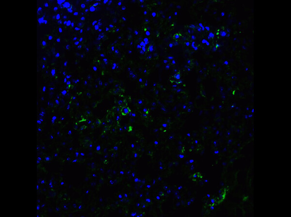
Immunofluorescence of Anti-ACE2 Antibody. Tissue: Rat Lung Tissue. Fixative: 4% PFA. Primary Antibody: Anti-ACE2 at 20µg/mL. Secondary Antibody: Goat anti-rabbit IgG secondary antibody at 1:500 dilution (green) and DAPI counterstain (blue).
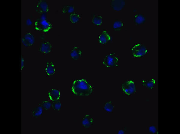
Immunofluorescence of Rabbit Anti-ACE2 Antibody. Tissue: Caco2 Cells. Fixative: 4%PFA. Primary Antibody: Anti-ACE2 at 5µg/mL. Secondary Antibody: Goat anti-rabbit IgG secondary antibody at 1:500 dilution (green) and DAPI counterstain (blue). Image showing membrane staining on Caco2 cells.
* VAT and and shipping costs not included. Errors and price changes excepted