
TACE Antibody, Rabbit, Polyclonal
Catalog Number:
ROC-600-401-H24
- Images (10)

| Article Name: | TACE Antibody, Rabbit, Polyclonal |
| Biozol Catalog Number: | ROC-600-401-H24 |
| Supplier Catalog Number: | 600-401-H24 |
| Alternative Catalog Number: | ROC-600-401-H24 |
| Manufacturer: | Rockland Immunochemicals |
| Host: | Rabbit |
| Category: | Antikörper |
| Application: | ELISA, IF, IHC, WB |
| Species Reactivity: | Human, Rat |
| Immunogen: | TACE Antibody was produced from whole rabbit serum prepared by repeated immunizations with a peptide corresponding to amino acids near the c-terminus of human TACE. This sequence differs from those of mouse and rat TACE by one amino acid. The immunogen is located within the last 50 amino acids of TACE. |
| Conjugation: | Unconjugated |
| Alternative Names: | ADAM17, ADAM18, CD156B, CSVP, NISBD, NISBD1, TACE |
| Application Dilute: | IF Microscopy: 10ug/ml, WB: 0.25-2µg/mL |
| Application Notes: | Anti-TACE Antibody is tested for use in ELISA, WB, ICC, and IF. Expect a band approximately ~93.0 kDa on specific lysates. 80 to 130kDa bands can be detected, which may represent mature protein, precursor and glycosylated TACE. Western Blot and Immunoflu |
|
|
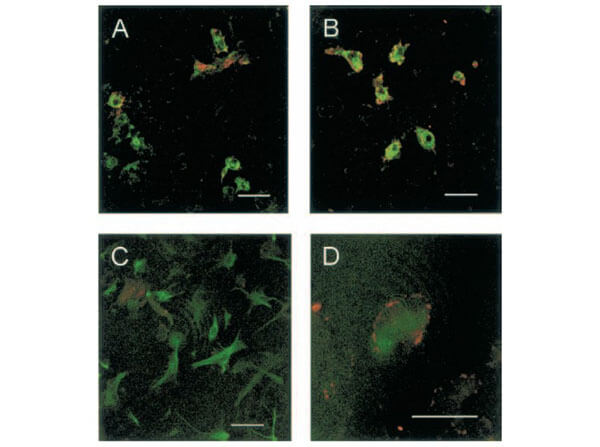
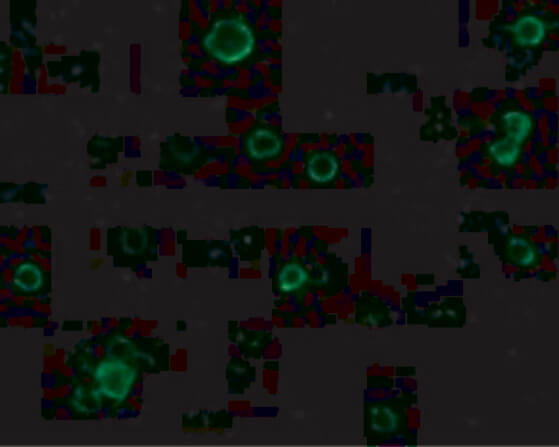
Immunofluorescence of Rabbit Anti-TACE Antibody. Cells: Rat Cortical Neurons. (A) Control cultures show TACE immunoreactivity at the cellular membrane of some microglial cells. (B) Glutamate-exposed cultures show that most microglial cells express TACE immunoreactivity. (C) Control cultures show that TACE immunostaining does not colocalize with astrocytes [glial fibrillary acidic protein (GFAP)-positive cells]. (D) Astrocyte (GFAP-positive cell) showing TACE immunoreactivity in its surface after treatment with glutamate. Double immunostaining of control and glutamate-exposed rat cortical cultures. (Hurtado et al., 2002). |
|
|
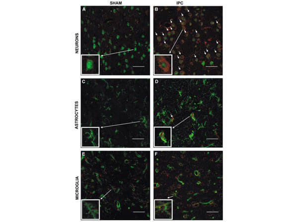
Immunofluorescence of Rabbit Anti-TACE Antibody. Cells: Rat Brain. Cellular localization of TACE. Double immunofluorescence staining of brain sections from sham-operated (SHAM, A, C, E) and IPC-exposed animals (IPC, B, D, F) of TACE (red) and the cellular markers (green) NeuN (neurons, A, B), GFAP (astrocytes, C, D) and L. esculentum lectin (microglia and endothelium, E, F). White arrows indicate TACE-positive cells. (Pradillo et al, 2005). |
|
|
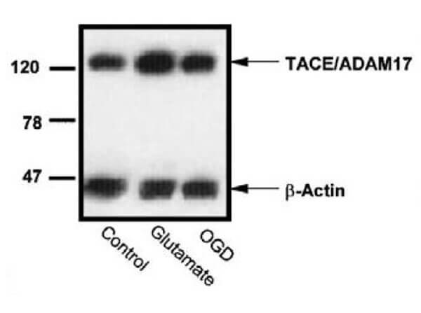
Western Blot of Anti-TACE Antibody. Effect of oxygen-glucose deprivation (OGD) or glutamate on the levels of TACE/ADAM17 in rat cortical cultures. (Hurtado et al., 2002). Western blot analysis of TACE in Rat Cortical Neuron homogenates from control, glutamate, and OGD-exposed cultures from a representative experiment. |
|
|
Western Blot of Rabbit Anti-TACE Antibody. Loading: 15 µg of lysates per lane. Primary Antibodies: Anti-TACE 600-401-H24 (0.5 µg/mL), TACE 22001 (2 µg/mL), and GAPDH (0.02 µg/mL), for 1h incubation at RT. Block: 5% NFDM/TBST. Secondary: Goat anti-rabbit IgG HRP conjugate at 1:10,000. Expect: ~93kDa. |
|
|
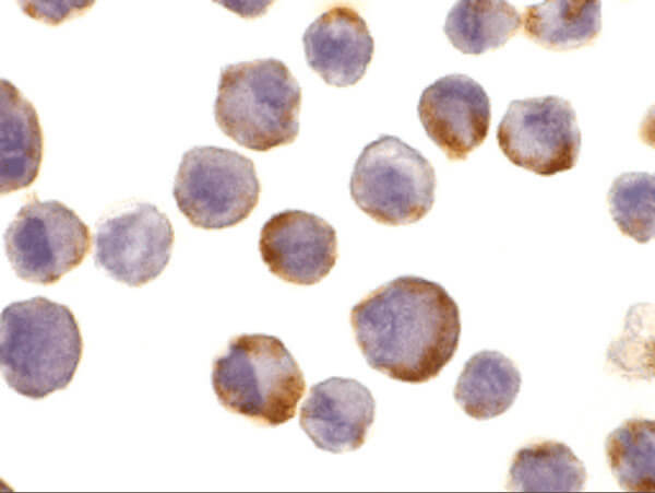
Immunocytochemistry of Rabbit Anti-TACE Antibody. Cells: HeLa cells. Primary Antibody: Anti-TACE antibody at 10 µg/ml overnight at 2-8C. Fixation: formaldehyde and blocked with 10% serum for 1 h at RT. Antigen Retrieval: heat mediation with a citrate buffer (pH6). Secondary Antibody: goat anti-rabbit IgG H&L (HRP) at 1:250. Counter stained with Hematoxylin. |
|
|
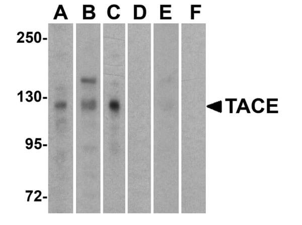
Western Blot of Rabbit Anti-TACE Antibody. Lane (A, D): HeLa. Lane (B, E): Jurkat. Lane (C, F): Raji. Lanes A-C are in the absence or Lanes D-F are in the presence of blocking peptide. Primary Antibody: Anti-TACE at 1µg/mL RT for 1hr. Secondary Antibody: Goat Anti-Rabbit IgG HRP at 1:10,000. Blocking 5% NFDM/TBST. Expect: ~93kDa. |
|
|
Immunofluorescence of Anti-TACE Antibody. Cells: HeLa Cells. Primary Antibody: Anti-TACE 10µg/mL. Secondary Antibody: Goat Anti-Rabbit IgG 1:500. Fixation: 4% paraformaldehyde. |
|
|
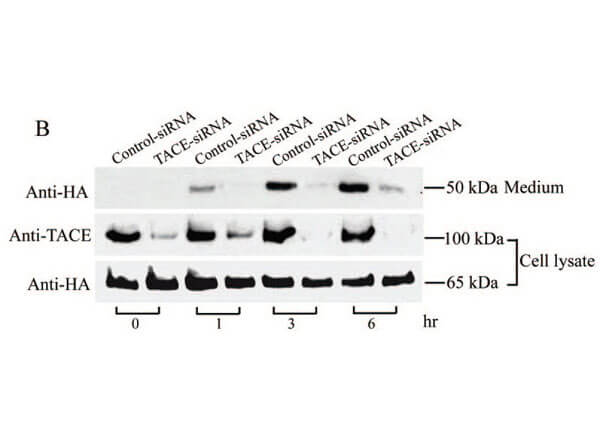
Western Blot of Rabbit Anti-TACE Antibody. Monkey COS cells stably expressing Pref-1A were transfected with control siRNA or TACE siRNA. TACE was detected in lysates by using the anti-TACE antibody. TACE expression levels were markedly reduced in TACE knockdown cell lysate. (Wang et al., 2006). |
|
|
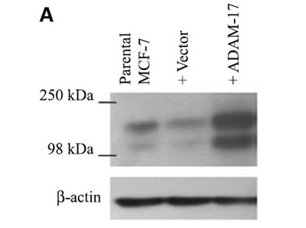
Western Blot of Anti-TACE (ADAM-17) Antibody. ADAM-17 (TACE) protein expression, following transfection of vector and ADAM-17 cDNA, was examined by immunoblot analysis with anti-ADAM-17 antibodies in MCF-7 cells. Increased ADAM-17 was detected in ADAM-17 transfected cells. (McGowan et al., 2007). |
|
|
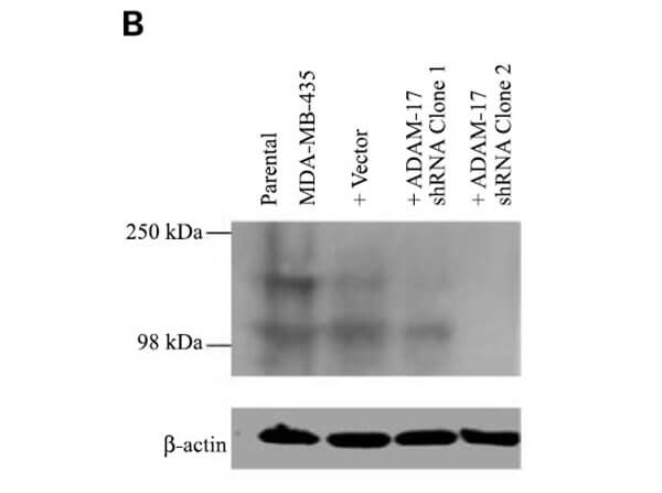
Western Blot of Rabbit Anti-TACE (ADAM-17). ADAM-17 (TACE) protein expression in MDA-MB-435 Cells, following transfection with ADAM-17 shRNA (two clones) or neomycin-resistant negative control vector, was examined by immunoblot analysis with anti-ADAM-17 antibodies. (McGowan et al., 2007). |
Product Guarantee and Expert Support
