
Histone H2A.Zac Antibody, Rabbit, Polyclonal
Catalog Number:
ROC-600-401-V36
- Images (7)

| Article Name: | Histone H2A.Zac Antibody, Rabbit, Polyclonal |
| Biozol Catalog Number: | ROC-600-401-V36 |
| Supplier Catalog Number: | 600-401-V36 |
| Alternative Catalog Number: | ROC-600-401-V36 |
| Manufacturer: | Rockland Immunochemicals |
| Host: | Rabbit |
| Category: | Antikörper |
| Application: | ChIP, DOT, ELISA, IF, WB |
| Species Reactivity: | Human, Mouse |
| Immunogen: | Anti-H2A.Zac Antibody was produced in rabbits by repeated immunization with a synthetic peptide containing the region of human histone H2A.Z acetylated at lysines 5, 7 and 11. |
| Conjugation: | Unconjugated |
| Alternative Names: | Histone H2A.Z, H2A/z |
| Application Dilute: | ELISA: 1:500, ChIP: 1 µg/ChIP, IF Microscopy: 1:500, WB: 1:1,000 |
| Application Notes: | Anti-H2A.Zac Antibody is tested for Chromatin Immunoprecipitation, Dot Blotting, ELISA, Immunofluorescence and Western Blots. Specific conditions for reactivity should be optimized by the end user. Expect a band approximately 15 kDa in the appropriate ce |
|
|
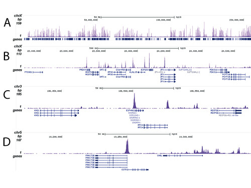
Chromatin Immunoprecipitation Rabbit Histone H2A.Zac Antibody. ChIP was performed on sheared chromatin from 100,000 human K562 cells using 1 µg the anti-H2A.Zac antiobdy. The 36 bp tags were aligned to the human genome using the ELAND algorithm. Figure A shows the peak distribution along the complete sequence, a 1.5 Mb region of the X-chromosome (figure B), and in two regions surrounding the EIF4A2 and CCT5 positive control genes, respectively (figure C and D). |
|
|
|
|
|
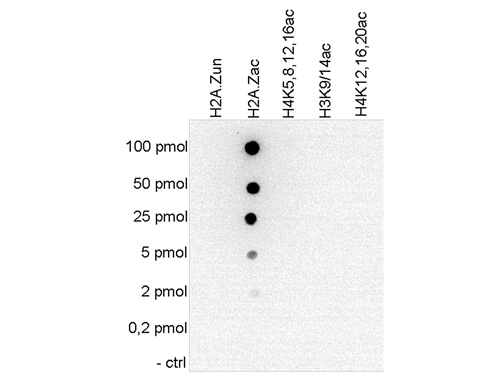
Dot Blot results of Rabbit anti-Histone H2 A.Zac antibody. Antigens: H2A.Z, H2A.Zac, H4K5,8,12,16ac, H3K9/14ac, H4K12,16,20ac. Load: 100pmol, 50pmol, 25pmol, 5pmol, 2pmol, 0.2pmol, and control. Primary antibody: Histone H2 A.Zac antibody at 1:20,000 overnight at 4C. Secondary antibody: anti-rabbit HRP secondary antibody at 1:10,000 for 45 min at RT. Block: 5% BLOTTO. |
|
|
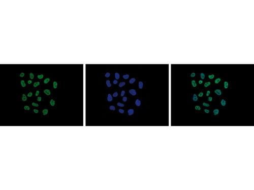
Immunofluorescence Microscopy results of Rabbit anti-Histone H2 A.Zac antibody. Tissue: HeLa cells. Fixation: 4% formaldehyde for 10 min. Block: PBS/Triton X-100 / 5% normal goat serum and 1% BSA. Primary antibody: Histone H2 A.Zac antibody at 1:500 for 1 hr at RT. Secondary antibody: anti-rabbit Alexa 488 secondary antibody at 1:10,000 for 45 min at RT. Staining: Histone H2 A.Zac antibody as green fluorescent signal (left), DAPI blue (middle), and a merge of the two stainings (right). |
|
|
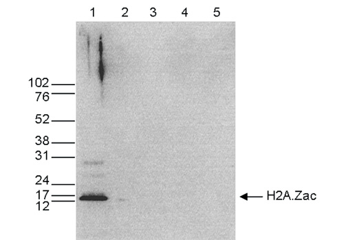
Western Blot results of Rabbit anti-Histone H2 A.Zac antibody. Lane 1: 25 µg HeLa whole cell extracts. Lane 2: 1µg recombinant histone H2A. Lane 3: 1µg recombinant histone H2B. Lane 4: 1µg recombinant histone H3. Lane 5: 1µg recombinant histone H4. Primary antibody: anti-Histone H2A.Zac antibody at 1:1,000 overnight at 4C. Secondary antibody: anti-rabbit HRP antibody at 1:10,000 for 45 min at RT. Block: TBS-Tween/5% BLOTTO. Predicted/Observed size: ~13.5 kDa for Histone H2 A.Zac. |
|
|
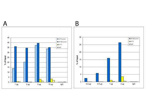
Chromatin Immunoprecipitation Rabbit Histone H2A.Zac Antibody. Figure A: ChIP using HeLa cells, Rabbit Histone H2A.Zac Antibody, and optimized PCR primer pairs for qPCR. ChIP was performed using sheared chromatin from 1,000,000 cells. A titration consisting of 1, 2, 5 and 10 µg of antibody per ChIP experiment was analyzed. IgG (2 µg/IP) was used as a negative IP control. Figure B: ChIP using human K562 cells, Rabbit Histone H2A.Zac Antibody, and optimized PCR primer sets for qPCR. ChIP was performed using sheared chromatin from 100,000 cells. A titration of the antibody consisting of 0.2, 0.5, 1 and 2 µg per ChIP experiment was analyzed. IgG (1 µg/IP) was used as negative IP control. qPCR was performed with primers specific for the promoter of the active genes CCT5 and EIF4A2, used as positive controls, and for the coding region of the inactive MYT1 gene and the Sat2 satellite repeat, used as negative controls. These figures shows the recovery, expressed as a % of input (the relative amount of immunoprecipitated DNA compared to input DNA after qPCR analysis). |
|
|
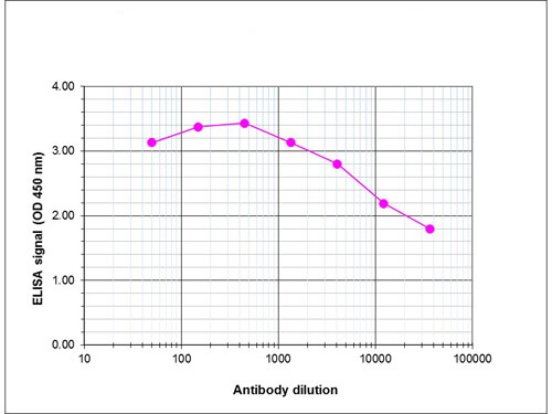
ELISA results of Rabbit anti-Histone H2 A.Zac antibody. Antigen: H2A.Zac peptide. Coating amount: 0.1 µg per well. Dilution series: serial. Mid-point concentration: 1:56,600 Histone H2 A.Zac antibody. Secondary antibody: Peroxidase anti-rabbit secondary antibody at 1:20,000. Substrate: TMB (p/n TMBE-1000). |
Product Guarantee and Expert Support
