The immunogen is a Red Fluorescent Protein (RFP) fusion protein corresponding to the full length amino acid sequence (234aa) derived from the mushroom polyp coral Discosoma.
Conjugation:
FITC
Alternative Names:
rabbit anti-RFP antibody fluorescein conjugation, FITC conjugated rabbit anti-RFP antibody, DsRed, rDsRed, Discosoma sp. Red Fluorescent Protein, Red fluorescent protein drFP583
0.02 M Potassium Phosphate, 0.15 M Sodium Chloride, pH 7.2
Form:
Lyophilized
Antibody Type:
Primary Antibody
Application Dilute:
FLISA: >1:20,000, IF Microscopy: 1:500 - 1:2,500, WB: >1:10,000
Application Notes:
Polyclonal anti-RFP is designed to detect RFP and its variants. This fluorescein conjugated antibody has been tested by dot blot and can be used to detect RFP by ELISA (sandwich or capture) for the direct binding of antigen. Significant amplification of
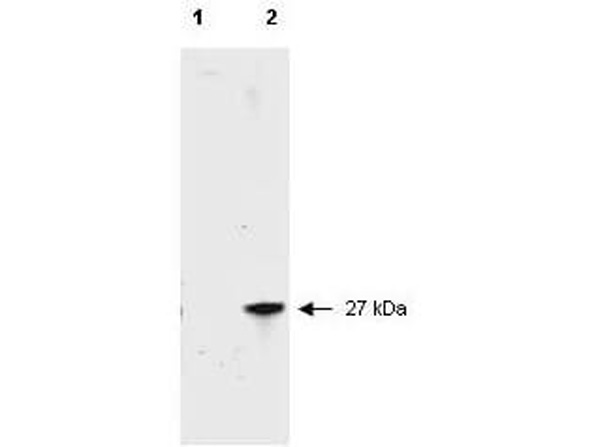
Western blot of RFP recombinant protein detected with Rocklands unconjugated polyclonal anti-RFP antibody (600-401-379). Lane 1 shows no reaction against a GFP recombinant protein present in 10 ug of HeLa cell extract. Lane 2 shows a single band detected in 10 ug of a HeLa lysate containing RFP recombinant protein. Rocklands polyclonal anti-RFP detects a 27 kDa band corresponding to the epitope tag RFP. A 4-12% Bis-Tris gradient gel (Invitrogen) was used for SDS-PAGE. The protein was transferred to nitrocellulose using standard methods. After blocking the membrane was probed with the unconjugated primary antibody diluted 1:2,500 for 1 h at room temperature followed by washes and reaction with a 1:5,000 dilution of IRDye(TM)800 conjugated Goat-a-Rabbit IgG [H&L] MX (611-132-122). IRDye(TM)800 fluorescence image was captured using the Odyssey Infrared Imaging System developed by LI-COR. IRDye is a trademark of LI-COR, Inc. Other detection systems will yield similar results.
* VAT and and shipping costs not included. Errors and price changes excepted