Collagen Type III Antibody Peroxidase Conjugated, HRP, Rabbit, Polyclonal
Biozol Catalog Number:
ROC-600-403-105
Supplier Catalog Number:
600-403-105
Alternative Catalog Number:
ROC-600-403-105
Manufacturer:
Rockland Immunochemicals
Host:
Rabbit
Category:
Antikörper
Species Reactivity:
Bovine, Human
Immunogen:
Collagen Type III from human and bovine placenta
Conjugation:
HRP
Alternative Names:
rabbit anti-Collagen Type III antibody peroxidase conjugation, HRP conjugated rabbit anti-Collagen Type III antibody, Collagen type III alpha 1 antibody, Collagen type III alpha antibody, EDS4A antibody, Ehlers Danlos syndrome type IV, autosomal dominant antibody, Fetal collagen antibody, COL3A1, Collagen alpha-1 (III) chain
Anti-Collagen Type III Peroxidase Conjugated Antibody is suitable for western blotting, IHC and for ELISA. Researchers should determine optimal titers for applications that are not stated below.
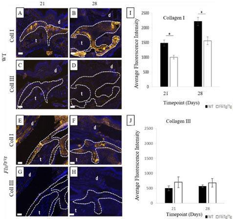
Immunohistochemistry of Anti-Collagen Type III Peroxidase Conjugated. WT mice have significantly increased collagen type I expression in the tendon adhesions. Representative images of collagen type I expression (gold) in the tendon adhesions of WT (a-b) andFliiTg/Tg(e-f) mice and collagen type III expression (gold) in the tendon adhesions of WT (c-d)FliiTg/Tg(g-h) at 21 and 28days post 50% partial laceration injury. (i) WT mice have significantly upregulated collagen type I levels in the adhesions compared withFliiTg/Tgmice. (j) No significant difference was noted in collagen type III levels in WT andFliiTg/Tgmice, although detectable levels of collagen type III were found in theFliiTg/Tgmice. DAPI is represented as blue fluorescence, t, tendon, d, dermis. Dotted line represents tendon adhesion area. Magnification * 10. Scale bar = 200µM and refers to all images. Data represented as mean SEM. *p 0.05.n= 6. Fig. 7. PMID: 32854733.
* VAT and and shipping costs not included. Errors and price changes excepted