The immunogen is a Red Fluorescent Protein (RFP) fusion protein corresponding to the full length amino acid sequence (234aa) derived from the mushroom polyp coral Discosoma.
Conjugation:
Biotin
Alternative Names:
chicken anti-RFP antibody biotin conjugation, biotin conjugated chicken anti-RFP antibody, DsRed, rDsRed, Discosoma sp. Red Fluorescent Protein, Red fluorescent protein drFP583
0.02 M Potassium Phosphate, 0.15 M Sodium Chloride, pH 7.2
Form:
Lyophilized
Antibody Type:
Primary Antibody
Application Dilute:
ELISA: 1:10,000, IF Microscopy: 1:100, WB: 1:2,000 - 1:5,000
Application Notes:
Anti-RFP is designed to detect recombinant RFP. This antibody has been tested to detect RFP by immunoblotting and immunofluorescence and is suitable for use in immunohistochemistry. Use either alkaline phosphatase or peroxidase conjugated polyclonal anti
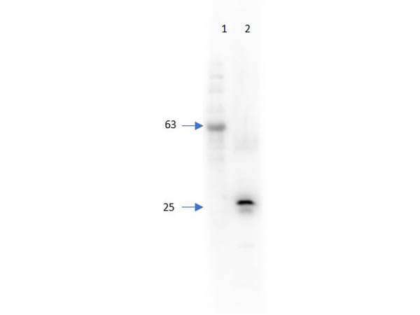
Western Blot of Chicken Anti-RFP Antibody Biotin Conjugated. Lane 1: Super Signal Molecular Weight. Lane 2: 50ng of RFP. Primary Antibody: Chicken Anti-RFP biotin conjugated at 1:1000 overnight at 2-8C. Secondary Antibody: HRP Streptavidin (p/n S000-03) 1:40,000 for 30mins at RT. Block: BlockOut Universal Blocking buffer (p/n MB-073). Expect: 27kDa.
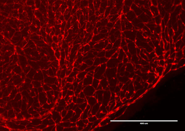
Immunofluorescence Microscopy of Chicken Anti-RFP antibody. Tissue: DsRed transgenic mouse retina. Fixation: 4% PFA. Blocking: 3% BSA, 0.3% Triton Primary antibody: RFP-biotin antibody at 1:100 for 12 h at 4C. Secondary antibody: Alexa488 secondary antibody at 1:10,000 for 4 hours at RT. Localization: RFP is nuclear and occasionally cytoplasmic. Staining: labeled in red.
* VAT and and shipping costs not included. Errors and price changes excepted