This product is designed for immunofluorescence microscopy, fluorescence based plate assays (FLISA) and fluorescent western blotting. This product is also suitable for multiplex analysis, including multicolor imaging, utilizing various commercial platfor
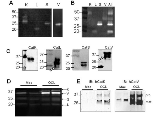
Western Blot results using Donkey Anti-Goat IgG FITC.Mature cathepsins K, L, S, and V are zymographically active and migrate at distinct electrophoretic distancesA) Recombinant cathepsins K, S, and V (1, 20, and 50 ng) from E. coli and cathepsin L (50 ng) isolated from human liver were loaded for cathepsin gelatin zymography and incubated overnight in acetate buffer, pH 6. The zymogram reveals zymographically active bands at different electrophoretic migration distances. B) Mature, recombinant cathepsins K, S, and V (10 ng) from eukaryotic expression systems and cathepsin L (50 ng) isolated from human liver were loaded separately and all in one lane (where indicated) for gelatin zymography assayed at pH 6. C) Western blot analysis of 50 ng of recombinant glycosylated cathepsin K, L, S, and V from eukaryotic expression systems also were loaded for non-reduced Western blotting. D) Monocyte-derived macrophages and monocyte-derived osteoclasts were lysed and equal amounts of protein were loaded for cathepsin zymography and E) reduced, fully denaturing Western blotting for cathepsins K and V. Procathepsin (pro) bands are at ~37 kDa and mature (mat) cathepsin bands are at ~27 kDa. Increased cathepsins K and V were detected in the osteoclasts compared to the macrophages. Figure 1. PMID: 21982919.
* VAT and and shipping costs not included. Errors and price changes excepted