0.02 M Potassium Phosphate, 0.15 M Sodium Chloride, pH 7.2
Form:
Lyophilized
Target:
Mouse
Antibody Type:
Secondary Antibody
Application Dilute:
FLISA: >1:20,000, IF Microscopy: >1:5,000, WB: >1:10,000
Application Notes:
Anti-Mouse IgG DyLight 488 Antibody has been tested by dot blot and western blot. This product is designed for immunofluorescence microscopy, fluorescence based plate assays (FLISA) and fluorescent western blotting. This product is also suitable for mult
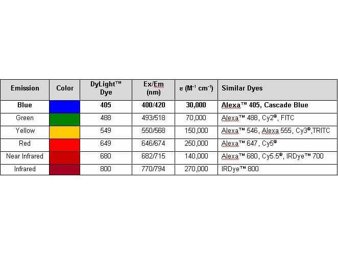
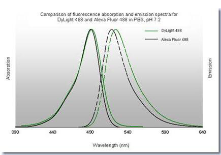
Properties of DyLight(TM) Fluorescent Dyes.
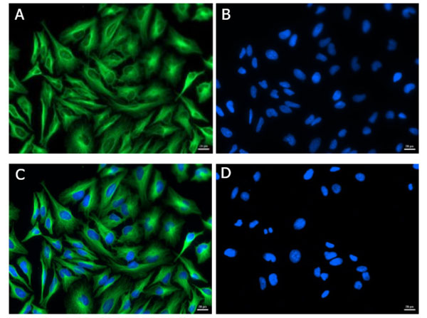
Immunofluorescence of Goat Anti-Mouse IgG (H&L) Antibody DyLight(TM) 488 Conjugated (Min X Bv Ch Gt GP Ham Hs Hu Rb Rt & Sh Serum Proteins). Cell line: HeLa. Primary Antibody: Alpha Tubulin (p/n 200-301-880) at 4 µg/mL (1:250) for 1hr at RT. Secondary Antibody: Goat Anti-Mouse DyLight(TM) 488 (p/n 610-141-121) at 1 µg/mL (1:1000) overnight at 4 C. Fixative: Ice Cold Methanol. Permeabilization: Ice Cold Methanol. Nuclear stain: Hoechst 33342. Expected Localization: Cytoplasmic. Image: A) Alpha Tubulin, B) Nuclear Stain, C) Merge, D) Secondary Only Control.
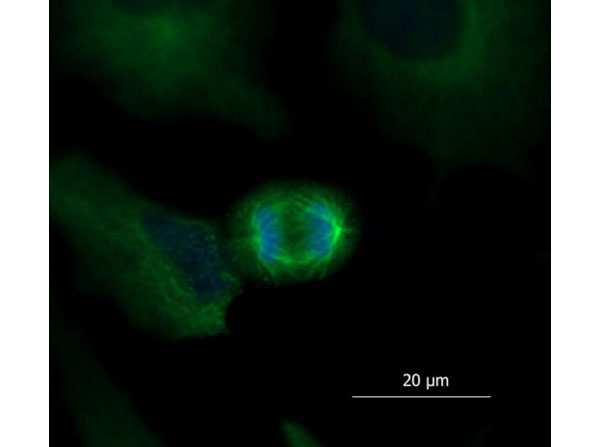
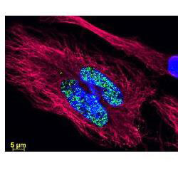
Immunofluorescence of Goat Anti-Mouse IgG (H&L) Antibody DyLight(TM) 488 Conjugated (Min X Bv Ch Gt GP Ham Hs Hu Rb Rt & Sh Serum Proteins). Cell line: HeLa. Primary Antibody: Alpha Tubulin (p/n 200-301-880) at 4 µg/mL (1:250) for 1hr at RT. Secondary Antibody: Goat Anti-Mouse DyLight(TM) 488 (p/n 610-141-121) at 0.1µg/mL (1:10000) overnight at 4 C. Fixative: Ice Cold Methanol. Permeabilization: Ice Cold Methanol. Nuclear stain: Hoechst 33342. Magnification: 40X. Expected Localization: Cytoplasmic. Image: HeLa cell nucleus in the anaphase stage of mitosis. Microtubule-based mitotic spindles are clearly visible.
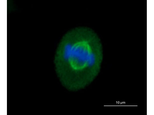
Immunofluorescence of Goat Anti-Mouse IgG (H&L) Antibody DyLight(TM) 488 Conjugated (Min X Bv Ch Gt GP Ham Hs Hu Rb Rt & Sh Serum Proteins). Cell line: HeLa. Primary Antibody: Alpha Tubulin (p/n 200-301-880) at 4 µg/mL (1:250) for 1hr at RT. Secondary Antibody: Goat Anti-Mouse DyLight(TM) 488 (p/n 610-141-121) at 0.1 µg/mL (1:10000) overnight at 4 C. Fixative: Ice Cold Methanol. Permeabilization: Ice Cold Methanol. Nuclear stain: Hoechst 33342. Magnification: 40X. Expected Localization: Cytoplasmic. Image: HeLa cell nucleus in the metaphase stage of mitosis. Microtubule-based mitotic spindles are clearly visible.
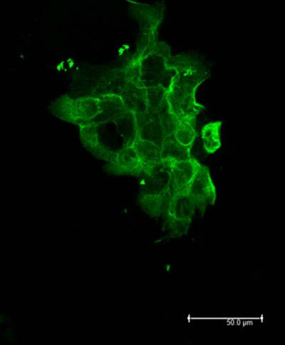
Rockland Dylight 488 Goat Anti Mouse IgG antibody-Immunofluorescence Cell Type: A431 cells Fixation: 4% paraformaldehyde 10 min Permeablization: 0.5% Triton X 30 min Primary Ab
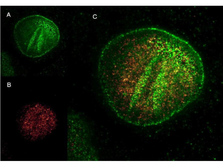
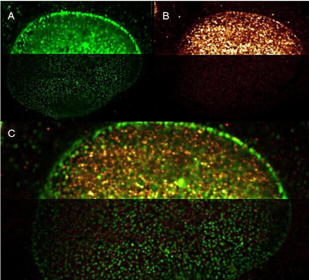
Rockland DyLight and ATTO dye conjugated antibodies provide high signal and low background for confocal microscopy and high resolution Stimulated Emission Depletion (STED) Microscopy. Both Dylight and Atto conjugated secondary antibodies maintained robust, intense signal during repeated laser excitation and de-excitation used during STED microscopy. Shown here are: A. (Green) Mouse anti NuP (NuP=Nuclear Pore Protein) detected with Dylight 488 Goat anti mouse (610-141-121) B. (Red) Rabbit Anti Ezh1/2 Pab (Ezh=enhancer of zeste homology) with detection by Rockland ATTO 425 conjugated Goat anti Rabbit (611-151-122) C. (Red and Green) Images combined. Data was collected on a STED-CW TCS-SP5 Confocal system (Leica Microsystems) equipped with a DFC 350FX Camera allowing sequential acquisition in wide-field, confocal and STED CW imaging modes and provided courtesy of: Myriam Gastard, PhD, personal communication, Leica Microsystems, Inc. USA
Rockland DyLight and ATTO dye conjugated antibodies provide high signal and low background for confocal microscopy (upper images) and high resolution Stimulated Emission Depletion (STED) Microscopy (lower images). Both Dylight and ATTO conjugated secondary antibodies maintained robust, intense signal during repeated laser excitation and de-excitation used during STED microscopy. Shown here are: A. (Green) Mouse anti NuP (NuP=Nuclear Pore Protein) detected with Dylight 488 Goat anti mouse (610-141-121) B. (Red) Rabbit Anti Ezh1/2 Pab (Ezh=enhancer of zeste homology) with detection by Rockland ATTO 425 conjugated Goat anti Rabbit (611-151-122) (Red and Green) Images combined. Data was collected on a STED-CW TCS-SP5 Confocal system (Leica Microsystems) equipped with a DFC 350FX Camera allowing sequential acquisition in widefield, confocal and STED CW imaging modes and provided courtesy of: Myriam Gastard, PhD, personal communication, Leica Microsystems, Inc. USA
* VAT and and shipping costs not included. Errors and price changes excepted