Mouse IgG Fc Antibody DyLight(TM) 649 Conjugated, Goat, Polyclonal
Biozol Catalog Number:
ROC-610-143-003
Supplier Catalog Number:
610-143-003
Alternative Catalog Number:
ROC-610-143-003
Manufacturer:
Rockland Immunochemicals
Host:
Goat
Category:
Antikörper
Species Reactivity:
Mouse
Immunogen:
Mouse IgG F(c) fragment
Conjugation:
DyLight 649
Alternative Names:
Goat Anti Mouse IgG F(c) Antibody DyLight(TM) 649 Conjugated, Goat Anti-Mouse IgG Fc Antibody DyLight(TM) 649 Conjugated, Goat Anti Mouse IgG Fc Fragment Antibody DyLight(TM) 649 Conjugated
Clonality:
Polyclonal
Concentration:
1.0 mg/mL by UV absorbance at 280 nm
Buffer:
0.02 M Potassium Phosphate, 0.15 M Sodium Chloride, pH 7.2
Form:
Lyophilized
Target:
Mouse
Antibody Type:
Secondary Antibody
Application Dilute:
FLISA: >1:20,000, IF Microscopy: >1:5,000, WB: >1:10,000
Application Notes:
The emission spectra for this DyLight(TM) conjugate match the principle output wavelengths of most common fluorescence instrumentation. This product is designed for immunofluorescence microscopy, fluorescence based plate assays (FLISA) and fluorescent weste
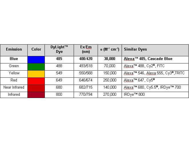
Properties of DyLight(TM) Conjugates.
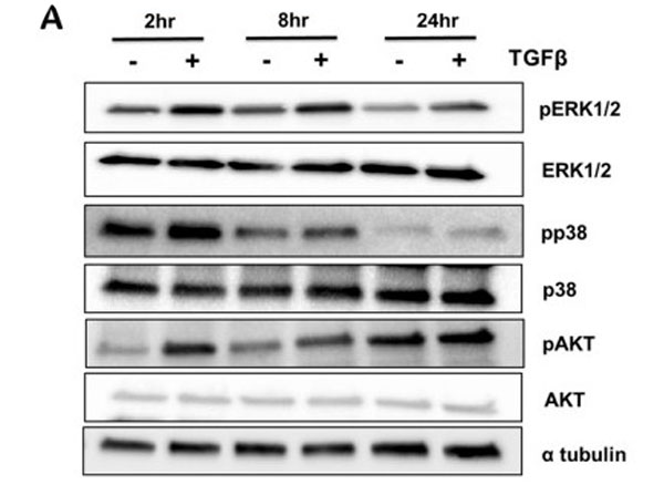
TGF-beta signaling regulates noncanonical pathways in the sclerotome. Sclerotome was treated with vehicle control or TGFbeta1 for 2, 8 or 24h. Immunoblot was used to determine activity of ERK, p38 and AKT. alpha tubulin was used as a general loading control. Immunoblots were cropped for clarity. Primary antibodies: pERK1/2 , ERK1/2, pp38, p38, pAKT, AKT, and Alpha tubulin [p/n 200-301-880] with secondary antibodies: anti-Rabbit-HRP and anti-mouse DyLight(TM)649 [p/n 610-143-003]. Figure 3. PMID: 33288795.
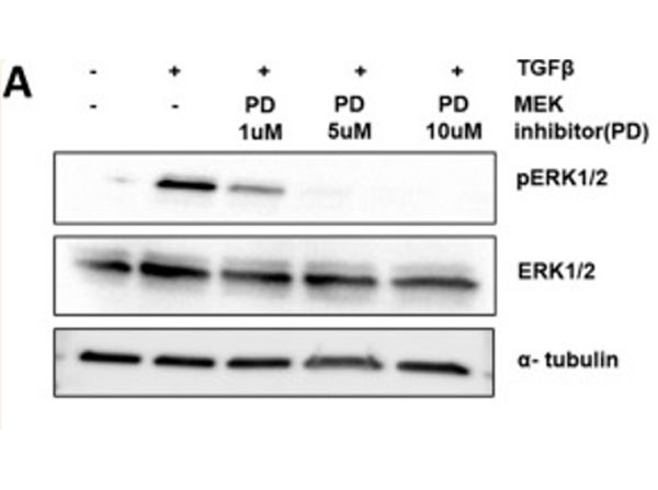
ERK is required for fibrous tissue marker regulation but not required to inhibit chondrogenesis. (A) Sclerotome was treated with a MEK inhibitor PD184352, PD, to inhibit ERK activity, for 24h and then cells were treated with TGFbeta1 for 8h. Immunoblot was used to determine relative levels of pERK1/2, ERK1/2, and alpha-tubulin. Primary antibodies: pERK1/2 , ERK1/2, and Alpha tubulin [p/n 200-301-880] with secondary antibodies: anti-Rabbit-HRP and anti-mouse DyLight(TM)649 [p/n 610-143-003]. Figure 5. PMID: 33288795.
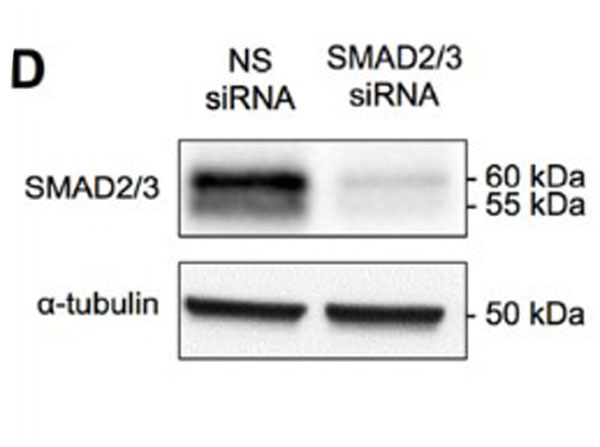
Knock-down ofSox9attenuates TGF-beta-mediated regulation ofPapss2 ATDC5 cells were transfected withSox9siRNA,Smad2/3siRNA, or control non-specific (NS) siRNA.Western blots showed Smad2/3 protein levels were reduced in the presence ofSmad2/3siRNA, alpha-Tubulin was used as a loading control (n = 6) (D).Primary antibodies: anti-SMAD2/3 antibody (1:2000) or anti-alpha-Tubulin antibody [1:2500, p/n 200-301-880], with secondary antibodies anti-mouse [1:2500, p/n 610-143-003]. Figure 4. PMID: 27746378.
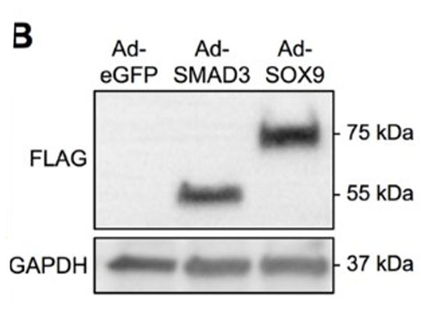
SMAD3 and SOX9 adenoviral vectors infect cells, induce expression of FLAG-tagged proteins, and up-regulate SMAD3 and SOX9 function respectively Bovine chondrocytes that were infected with either Ad-eGFP, Ad-SMAD3, or Ad-SOX9. Western blots of extracts from bovine chondrocytes showed expression of FLAG-tagged proteins at approximately 52 kDa and 75 kDa, corresponding to SMAD3 and SOX9 molecular weights respectively (n = 4) (B). Primary antibodies: anti-GAPDH antibody (1:1000) or anti-FLAG antibody (1:500), with secondary antibodies: HRP-conjugated anti-rabbit (1:2000) and anti-mouse [1:2500, p/n 610-143-003] Figure 2. PMID: 27746378.
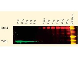
DyLight(TM) dyes can be used for two-color Western Blot detection with low background and high signal. Anti-tubulin was detected using a DyLight(TM) 549 conjugate. Anti-TNFa was detected using a DyLight(TM) 649 conjugate. The image was captured using the Typhoon(TM) 9410 Imaging System.
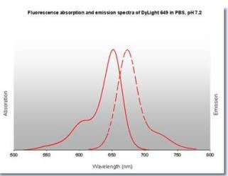
DyLight(TM) 649 Fluorescence Spectra.
* VAT and and shipping costs not included. Errors and price changes excepted