0.02 M Potassium Phosphate, 0.15 M Sodium Chloride, pH 7.2
Form:
Lyophilized
Target:
Mouse
Antibody Type:
Secondary Antibody
Application Dilute:
FLISA: >1:20,000, IF Microscopy: >1:5,000, WB: >1:10,000
Application Notes:
Anti-Mouse IgG DyLight 800 Antibody has been tested by dot blot and western blot and is designed for immunofluorescence microscopy, fluorescence based plate assays (FLISA) and fluorescent western blotting. This product is also suitable for multiplex anal
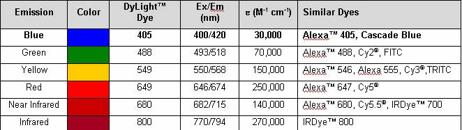
Properties of DyLight(TM) Fluorescent Dyes.
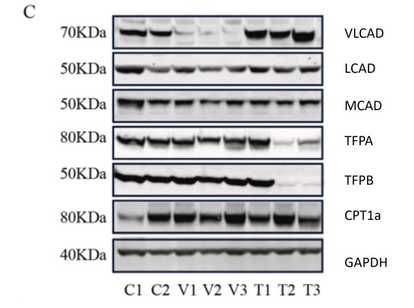
C. Representative western blots, original blots are shown in (supplementary Fig S8-9). And densitometric quantification of relative protein levels from western blots. Data are depicted as mean SD, n = 3, **P < 0.01, ***P < 0.001 and ****P < 0.0001 by one-way ANOVA. Intracellular transport, activation, mitochondrial transport, beta-oxidation, carnitine shuttle, and auxiliary proteins. The primary antibodies used as follows: VLCAD 1:1000, MCAD 1:1000, LCAD 1:1000, TFPa 1:500, TFPb 1:3000, CPT1alpha 1:1000, and GAPDH 1:30,000 dilutions overnight at 4 C. The membranes were then incubated with fluorescent conjugated secondary antibodies for 1 h, DyLight 800 conjugated goat Anti-Rabbit IgG (611-145-002), DyLight 680 conjugated goat Anti-Rabbit IgG (611-144-003), DyLight 800 conjugated goat Anti-Mouse IgG (610-145-002), and DyLight 680 conjugated donkey Anti-Mouse IgG (610-744-124). Fig 1. PMID: 33725513.
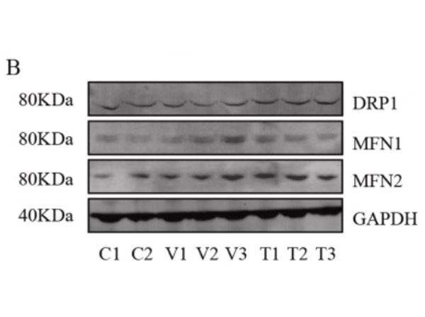
Assessment of mitochondrial fusion and fission. B. Representative western blots (original blots are shown in supplementary Fig. S10) and quantification of MFN1/2 and DRP1. No significant changes in the relative levels of proteins that facilitate mitochondrial fusion (MFN1/2) and fission (DRP1) between non-disease (control) and mutant primary fibroblasts. Data are depicted as mean SD, n = 3. The primary antibodies used as follows: MFN1 1:400, MFN2 ( 1:400, DRP1 1:100 and GAPDH 1:30,000 dilutions overnight at 4 C. The membranes were then incubated with fluorescent conjugated secondary antibodies for 1 h, DyLight 800 conjugated goat Anti-Rabbit IgG (611-145-002), Antibody DyLight 680 conjugated Anti-Rabbit IgG made in goat (611-144-003), DyLight 800 conjugated goat Anti-Mouse IgG (610-145-002), and DyLight 680 conjugated donkey Anti-Mouse IgG (610-744-124). Fig 3. PMID: 33725513.
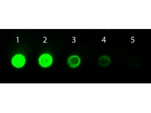
Dot Blot of Goat anti-Mouse IgG Antibody DyLight 800 Conjugated. Antigen: Mouse IgG. Load: Lane 1 - 100 ng Lane 2 - 33.3 ng Lane 3 - 11.1 ng Lane 4 - 3.70 ng Lane 5 - 1.23 ng. Primary antibody: none. Secondary antibody: Goat anti-Mouse IgG Antibody DyLight 800 Conjugated at 1:1,000 for 60 min at RT. Block: MB-070 for 60 min at RT.
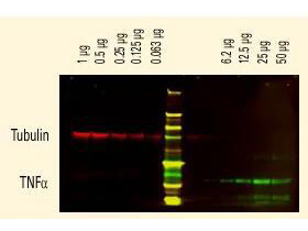
DyLight(TM) dyes can be used for two-color Western Blot detection with low background and high signal. Anti-tubulin was detected using a DyLight(TM) 680 conjugate. Anti-TNFa was detected using a DyLight(TM) 800 conjugate. The image was captured using the Odyssey Infrared Imaging System developed by LI-COR.
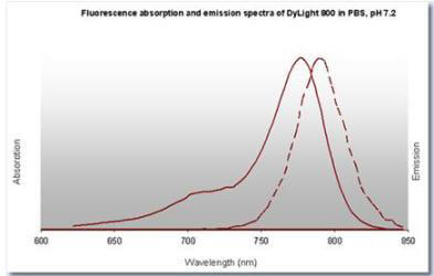
DyLight(TM) 800 Fluorescence Spectra
* VAT and and shipping costs not included. Errors and price changes excepted