0.02 M Potassium Phosphate, 0.15 M Sodium Chloride, pH 7.2
Form:
Lyophilized
Target:
Rabbit
Antibody Type:
Secondary Antibody
Application Dilute:
FLISA: >1:20,000, IF Microscopy: >1:5,000, WB: >1:10,000-1:25,000
Application Notes:
Anti-Rabbit IgG (H&L) DyLight 488 Antibody has been tested by western blot and is designed for immunofluorescence microscopy, fluorescence based plate assays (FLISA) and fluorescent western blotting. This product is also suitable for multiplex analysis,
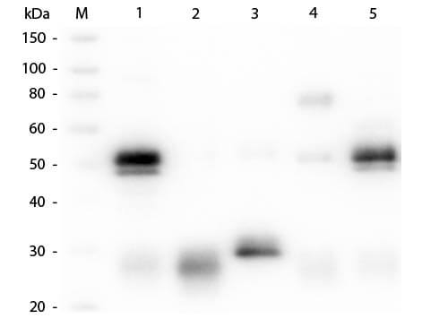
Western Blot of Unconjugated Anti-Rabbit IgG (H&L) (GOAT) Antibody (Min X Bv, Ch, Gt, GP, Ham, Hs, Hu, Ms, Rt & Sh Serum Proteins) (p/n 611-101-122). Lane M: 3 µl Molecular Ladder. Lane 1: Rabbit IgG whole molecule (p/n 011-0102). Lane 2: Rabbit IgG F(ab) Fragment (p/n 011-0105). Lane 3: Rabbit IgG F(c) Fragment (p/n 010-0103). Lane 4: Rabbit IgM Whole Molecule (p/n 011-0107). Lane 5: Normal Rabbit Serum (p/n B309). All samples were reduced. Load: 50 ng per lane. Block: MB-070 for 30 min at RT. Primary Antibody: Anti-Rabbit IgG (H&L) (GOAT) Antibody (Min X Bv, Ch, Gt, GP, Ham, Hs, Hu, Ms, Rt & Sh Serum Proteins) (p/n 611-101-122) 1:1,000 for 60 min at RT. Secondary antibody: Anti-Goat IgG (DONKEY) Peroxidase Conjugated Antibody (p/n CUST10) 1:40,000 in MB-070 for 30 min at RT. Predicted/Observed Size: 25 and 50 kDa for Rabbit IgG and Serum, 25 kDa for F(c) and F(ab), 70 and 23 kDa for IgM. Rabbit F(c) migrates slightly higher.
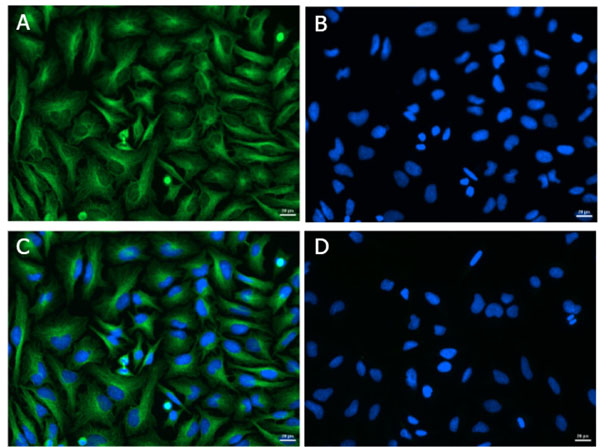
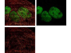
Immunofluorescence of Goat Anti-Rabbit IgG (H&L) Antibody DyLight(TM) 488 Conjugated (Min X Bv Ch Gt GP Ham Hs Hu Ms Rt & Sh Serum Proteins). Cell line: HeLa. Primary Antibody: Alpha Tubulin (p/n 600-401-880) at 4.4 µg/mL (1:250) for 1hr at RT. Secondary Antibody: Goat Anti-Rabbit DyLight(TM) 488 (p/n 611-141-122) at 1 µg/mL (1:1000) overnight at 4 C. Fixative: Ice Cold Methanol. Permeabilization: Ice Cold Methanol. Nuclear stain: Hoechst 33342. Expected Localization: Cytoplasmic. Image: A) Alpha Tubulin, B) Nuclear Stain, C) Merge, D) Secondary Only Control.
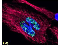
DyLight(TM) dyes can be used for multi-color immunofluorescence microscopy with uniform fluorescence intensity throughout the image. DyLight(TM) dyes are exceptionally bright and photostable and are optimized for microscopy and microarray detection methods. This image shows anti-histone detection using a DyLight(TM) 488 conjugate (green). Anti-Tubulin was detected using a DyLight(TM) 549 conjugate (red). Nuclei were counter-stained using DAPI (blue). The image was captured using an Axio Imager.Z1 (Zeiss Micro Imaging Inc).
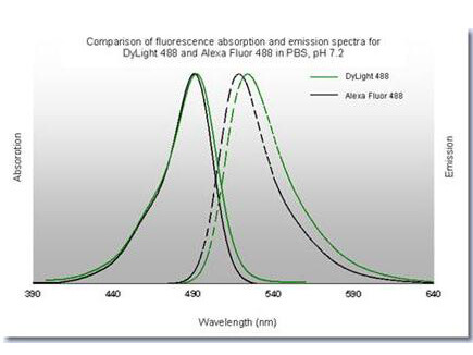
DyLight(TM) 488 Fluorescence Spectra.
DyLight(TM) 488 conjugated anti-Rabbit IgG was used to demonstrate 2 color STED immunofluorescence microscopy. Methanol fixed A431 cells were blocked with normal goat serum. The cells were then probed with 0.4 µg/mL final concentration of anti-HDAC and detected with 0.2 µg/mL DyLight(TM)488 conjugated Anti-RABBIT IgG [GOAT] secondary antibody (colored GREEN). Also shown in this 2-color STED image is Rocklands a-tubulin monoclonal antibody [MOUSE] (p/n 200-301-880) detected with ATTO 425 conjugated anti-MOUSE IgG [GOAT] (610-151-121) secondary antibody (colored RED). Image courtesy of Myriam Gastard, Leica Microsystems, USA.
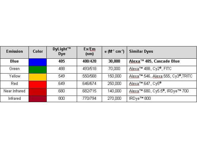
Properties of DyLight(TM) Fluorescent Dyes.
* VAT and and shipping costs not included. Errors and price changes excepted