This product is designed for immunofluorescence microscopy, fluorescence based plate assays (FLISA) and fluorescent western blotting. This product is also suitable for multiplex analysis, including multicolor imaging, utilizing various commercial platfor
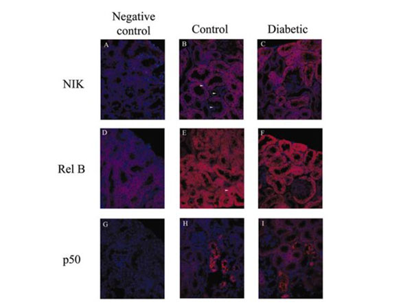
Representative 5-µm formalin-fixed sections of kidney sampled from control (B,E, andH) and diabetic (C,F, andI) mice. Negative controls (eliminating the primary antibody) are shown for the diabetic tissues inA,D, andG. Secondary antibody used for both NIK and RelB was Texas Red-conjugated antibody. While NIK was predominantly located in proximal tubular epithelial cells in controls and diabetics, RelB staining was distributed throughout all tubules in the cortex. Little immunostaining was observed in the glomeruli for NIK and RelB. p50 immunostaining was localized to only a few tubules in each section of control and diabetic kidneys. *400 magnification. Fig 6. PMID: 16644679.
* VAT and and shipping costs not included. Errors and price changes excepted