![]()
|
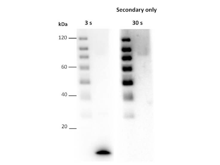
Western blot detection using Chemiluminescent FemtoMax(TM) HRP Substrate. rPARP1 domain detected at 11 kDa after 3 sec exposure using primary antibody rabbit anti-serum 1:500 overnight, at 4C. Secondary antibody Peroxidase Goat Anti-Rabbit IgG Antibody at 1:40,000. All incubations were performed in Blocking Buffer for Fluorescent Western Blocking (p/n MB-070). |
![]()
|
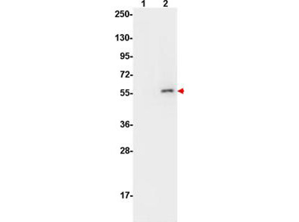
Western Blot of Mouse Anti-AKT pS473 antibody using Femtomax. Lane 1: non-phosphorylated AKT in untreated cells. Lane 2: phosphorylated AKT (indicated by arrowhead at ~56 kDa) on PDGF stimulated NIH/3T3 cell lysates. Load: 10 µg per lane. Primary antibody: AKT pS473 antibody at 1:10,000 in TBS with 0.05% Tween-20 with 1% BSA, for 1 h at 4 C. Secondary antibody: HRP conjugated Gt-a-Mouse IgG (p/n 610-103-121) was used at a 1:20,000 dilution for 1 h at 4 C with Chemiluminescent FemtoMax(TM) Super Sensitive HRP Substrate (p/n FEMTOMAX-100). |
![]()
|
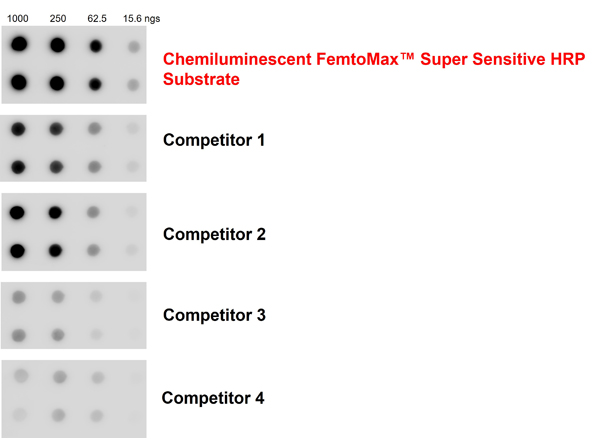
Chemiluminescent FemtoMax(TM) Super Sensitive HRP Substrate and competitor comparison. GAPDH protein was dotted on a nitrocellulose membrane, load 1000, 250, 62.5, 15.6ngs. The membrane was blocked for one hour at room temperature. Primary antibody: Rb anti-GAPDH diluted 1:2000 and incubated at 4C overnight. After washing, secondary antibody: Gt anti-Rb IgG-HRP diluted 1:50,000 at room temperature for 2 hours. Detection: Chemiluminescent FemtoMax(TM) Super Sensitive HRP Substrate or competitor 1-4. |
![]()
|
Chemiluminescent FemtoMax(TM) Super Sensitive HRP Substrate for Microwell and/or Membrane (2 component system) |
![]()
|
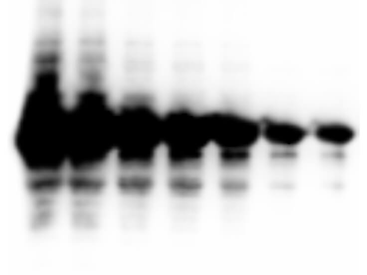
Western Blot of anti-GST tag antibody. Lane 1: Recombinant GST tagged recombinant protein 5 ug. Lane 2: Recombinant GST tagged recombinant protein 2 ug. Lane 3: Recombinant GST tagged recombinant protein 1 ug. Lane 4: Recombinant GST tagged recombinant protein 500 ng. Lane 5: Recombinant GST tagged recombinant protein 250 ng. Lane 6: Recombinant GST tagged recombinant protein 100 ng. Lane 7: Recombinant GST tagged recombinant protein 50 ng. Primary antibody: anti-GST antibody at 1:1000 for overnight at 4C. Secondary antibody: donkey secondary antibody at 1:10,000 for 45 min at RT. Block: 5% BLOTTO overnight at 4C. Predicted/Observed size: 78 kDa. |
![]()
|
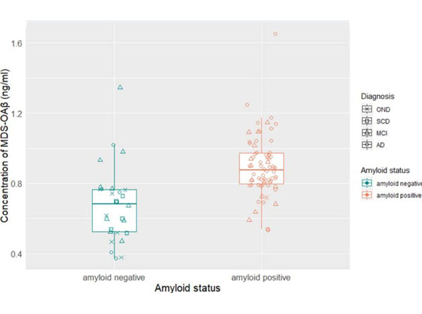
Concentration of plasma MDS-OAbeta according to groups.The incubated plasma sample mixture and serially diluted standard samples were added to respective wells, and the plates were incubated at room temperature for 1 hour. Afterwards, 100 µL/well of enhanced chemiluminescence substrate solution (p/n Femtomax) was added, and the Relative Luminescence Unit (RLU) signal was detected using a Victor 3TMmulti-spectrophotometer.Abbreviations:AD, Alzheimers disease, MCI, mild cognitive impairment, MDS-OAbeta, Multimer Detection System-Oligomeric Amyloid-beta, OND, other neurodegenerative disease, SCD, subjective cognitive decline.Fig 1. PMID: 33958861. |
![]()
|
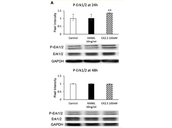
Effect of CK2.3 on p-Erk 1/2 in RANKL-induced osteoclastogenesis. CK2.3 increased p-Erk 1/2 after 24 h of stimulation in RAW264.7 cells, as determined by (A) Western blotting. The blot was incubated in 3% BSA for 1 h to block non-specific binding. Antibodies used at 1:1000 dilutions (in 1% BSA) overnight at 4 C. Followed by incubation with the secondary antibody HRP anti-rabbit at a 1:5000 dilution (in 1% BSA). The blot was incubated in Chemiluminescent FemtoMax Super Sensitive HRP Substrate (p/n Femtomax) for 2 min. Fig 6. PMID: 32660129. |
![]()
|
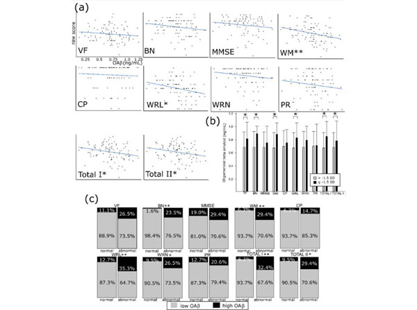
The subtests of the Korean version of Consortium to Establish a Registry for Alzheimers disease (CERAD-K) and plasma oligomerized beta amyloid (OAbeta). (a) Raw scores of the CERAD-K and OAbeta concentration. (b) The abnormal CERAD group (below -1.5 standard deviation of age/sex/education adjusting norms) in verbal fluency, naming, word memory/recall, and total scores showed higher OAbeta concentration compared with control. (c) Abnormality in naming, word memory/recall/recognition, and total scores are significantly more frequent in high OAbeta groups ( 0.78 ng/mL). SD-standard deviation, VF-ve |