![]()
|
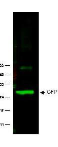
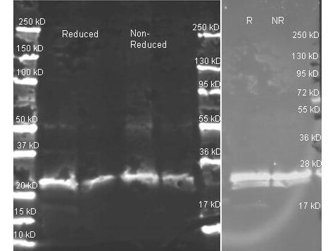
Rockland anti GFP polyclonal antibody (600-401-215) was used to detect GFP protein. Wild type GFP (0.1 µg) was used to spike 30 µg of a HeLa whole cell lysate. This antibody detects a 27 kDa band corresponding to the epitope tag GFP. A 4-20% Tris-Glycine gradient gel was used for SDS-PAGE. The protein was transferred to nitrocellulose using standard methods. After blocking with 5% BLOTTO in PBS, the membrane was probed overnight at 4 C with the primary antibody diluted in 5% BLOTTO to 1:1,000, followed by washes and reaction with a 1:10,000 dilution of IRDye 800 conjugated Goat-a-Rabbit IgG [H&L] MX10 (611-132-122). IRDye 800 fluorescence image was captured using the Odyssey Infrared Imaging System developed by LI-COR. IRDye is a trademark of LI-COR, Inc. Other detection systems will yield similar results. |
![]()
|
|
![]()
|
This product is assembled as a kit. See attached protocol or CofA for further details. |
![]()
|
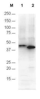
Anti-6X His epitope tag polyclonal antibody (600-401-382) detects His-tagged recombinant proteins by western blot. The blot was blocked with 3% BSA in TBST for 45 min at RT. Antibody was incubated with blot at a 1:1,000 dilution in TBST with 3% BSA for 1 hour at RT. Detection occurred using HRP Gt-a-Rabbit IgG (p/n 611-103-127) diluted 1:80,000 in blocking buffer (p/n MB-070) for 30 min at RT. Lane 1 was loaded with 12-Epitope Tag Protein Marker Lysate (p/n MB-301-0100) which has the His epitope tag incorporated through a C-terminal linkage (~42 kDa). Lane 2 was loaded with His-SUMO-GFP recombinant protein which has the His epitope tag incorporated through an N-terminal linkage (~40 kDa). A 4-20% gradient gel was used to resolve the protein by SDS-PAGE. Proteins were transferred to nitrocellulose using standard methods. Molecular weights were estimated by comparison to standards (lane M). |
![]()
|
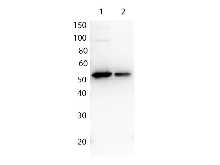
Rockland Affinity Purified anti FLAG(TM) Polyclonal Antibody (600-401-383) detects both C terminal linked and N terminal linked FLAG(TM) tagged recombinant proteins by western blot. This antibody was used at a dilution of 1:2,500 to detect 1.0 ?g of recombinant protein containing either the FLAG(TM) epitope tag linked at the carboxy (C) or the amino (N) terminus of the recombinant protein. A 4-20% gradient gel was used to resolve the protein by SDS-PAGE. The protein was transferred to nitrocellulose using standard methods. After blocking, the membrane was probed with the primary antibody for 1 h at room temperature followed by washes and reaction with a 1:10,000 dilution of IRDye 800 conjugated Gt-a-Rabbit IgG (H&L) MX10 (code 611-132-122) for 30 min at room temperature. LICORs Odyssey Infrared Imaging System was used to scan and process the image. Other detection systems will yield similar results. |
![]()
|
Rocklands anti-GST polyclonal antibody (600-101-200) in western blot shows detection of recombinant GST (indicated by band at ~ 28 kDa). The SDS-PAGE contained approximately 0.2 µg of rGST loaded on to a 4-20% gradient gel for separation. After electrophoresis, the gel was transferred to nitrocellulose and blocked with ''Blocking Buffer for Fluorescent Western Blotting p/n MB-070 in TBS for 1h at RT. The membrane was probed with anti-GST antibody at a 1:2,000 dilution in blocking reagent, overnight at 4 C. For detection DyLight(TM)800 conjugated Donkey-a-Goat IgG (p/n 605-745-002) was used at a 1:20,000 dilution (in blocking reagent) for 30 min at 25 C. Fluorescent data was collected on a LICOR Odyssey instrument. |
![]()
|
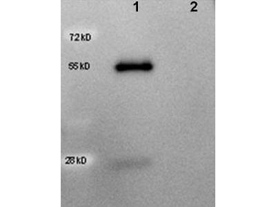
Western Blot of HRP anti-Goat IgG antibody (605-4313) showing detection of 50ng of goat IgG (lane 1) but not human IgG (lane 2). Samples were separated by 4-20% SDS-PAGE under reducing conditions and transferred to nitrocellulose membrane. The blot was blocked overnight at 4 C in 5% BSA in TBS. A 1:5,000 dilution of antibody in Blocking Buffer for Fluorescent Western Blotting (p/n MB-070) was used to probe the membrane at room temperature for 1 h. The image was developed using Chemiluminescent FemtoMax(TM) Super Sensitive HRP Subs |