Multiplex Blocking Buffer, Fluorescent Blocking Buffer, Blocking Solution, Blocking Buffer Western Blot, IRDye Western Blot Blocking Buffer, Alexa Dye Blocking Buffer, DyLight Blocking Buffer
Concentration:
1X
Buffer:
See application note.
Form:
Liquid (sterile filtered)
Application Dilute:
WB: User Defined
Application Notes:
Fluorescence technology is widely used to detect proteins in both the visible and near-infrared ranges. This product allows for superior signal detection and lower background noise when fluorochrome conjugated antibodies are used to visualize proteins in
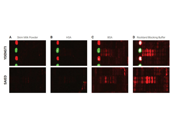
Selected sections of the PEPperCHIP Peptide Microarrays after assay with different blocking reagents. The microarrays were blocked for 30 minutes with either 2% skim milk powder (A), 1% HSA (B), 1% BSA (C) or 100% Rockland Blocking Buffer [p/n MB-070] (D), respectively. A human serum sample was assayed at dilution 1:200, followed by detection with secondary goat Anti-Human IgG (H+L) DyLight(TM) 680 Antibody [p/n 609-144-123]. Red spots = sample responses and polio control peptides, green spots = HA control peptides. The underlying binding motifs of the respective sections are indicated on the left.
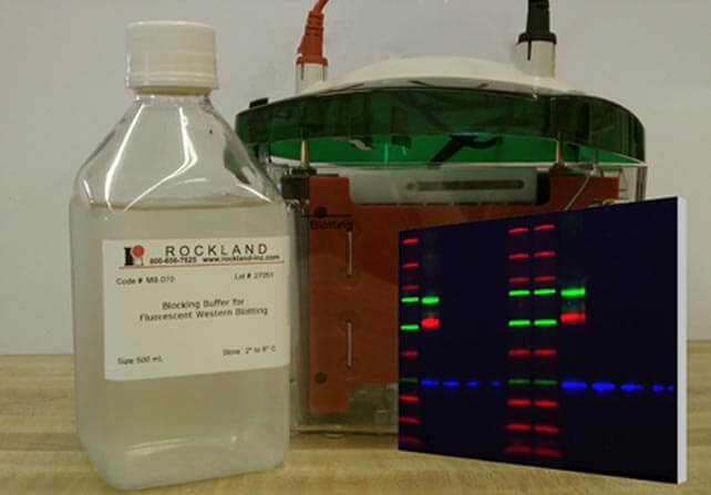
Blocking Buffer for Fluorescent Western Blotting
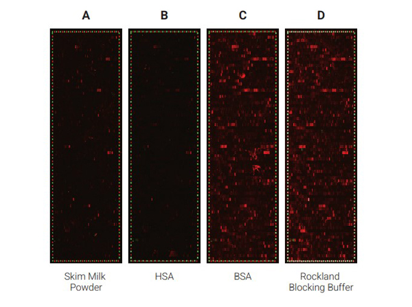
Comparison of the performance of different blocking reagents in epitope mappings with PEPperCHIP Peptide Microarrays.The PEPperCHIP Peptide Microarrays were blocked for 30 minutes with either 2% skim milk powder (A), 1% HSA (B), 1% BSA (C) or 100% Rockland Blocking Buffer [p/n MB-070] (D). A human serum sample was assayed at dilution 1:200, followed by detection with secondary goat anti-Human IgG (H+L) DyLight(TM) 680 Antibody [p/n 609-144-123] and a control anti-HA (12CA5)-DyLight(TM) 800 Antibody. Red spots = sample IgG response and frame of polio control peptides, green spots = frame of HA control peptides.
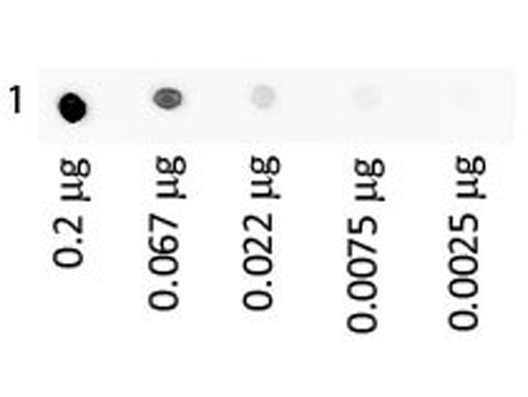
Dot Blot of Human IgA Fluorescein using MB-070. Antigen: Human IgA Fluorescein. Load: 3-fold serial dilution starting at 200 ng. Block: MB-070 for 30 min at RT.
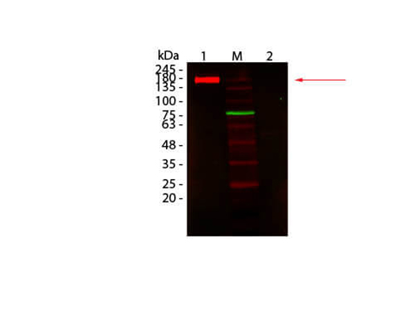
Western Blot of Fluorescent TrueBlot: Anti-Rabbit IgG DyLight 680 Conjugated using MB-070. Lane 1: Rabbit IgG, Non-denatured. Lane 2: Rabbit IgG, Denatured. Load: 50 ng per lane. Primary antibody: none. Secondary antibody: Fluorescent TrueBlot: Anti-Rabbit IgG DyLight 680 Conjugated antibody at 1:1,000 for 60 min at RT. Block: MB-070 for 30 min at RT. Predicted: 160 kDa for non-denatured, observed: 170-180 kDa for non-denatured. Band migrates at slightly higher molecular weight.
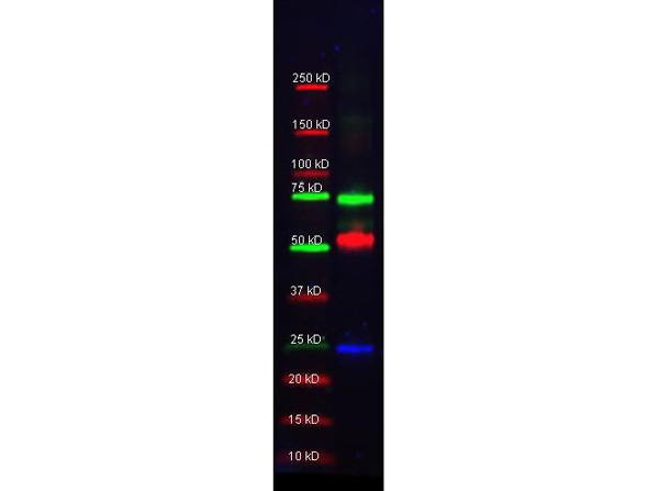
Multiplex western blot results using MB-070. Rockland Mouse-a-GST (200-301-200 lot 24882, blue), Rabbit anti-Transferrin (109-4134 lot 3033), and Goat-anti-Alpha-1-Anti-Trypsin (100-101-147 lot 5842) were used in a multiplex system to detect target proteins under reducing conditions in albumin depleted human serum with 320 ng of added GST. Sample was run by SDS-PAGE, transferred to 0.2 um PVDF using the BioRad Trans-Blot Turbo and blocked in 2.5% Blotto, 2.5% BSA, 0.02% Tween over night at 4C. Membrane was probed with three primary antibodies at 1:1000 dilution (in MB-070 over night at 4C). Detection shown was using DyLight(TM)549 Donkey anti-Rabbit IgG (611-742-127 lot 21100, shown as green) DyLight(TM)488 Donkey anti-Mouse IgG (610-741-124 lot 21095, shown as blue), and DyLight(TM)649 Donkey anti-Goat IgG (605-743-125 lot 20834, shown as red) at 1:10,000 (in MB-070 at 30 min RT). Blots were washed, rinsed in methanol, dried and Images were collected using the BioRad VersaDoc System.
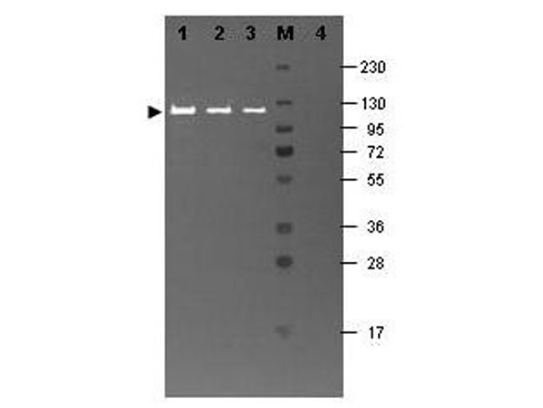
Western blot results using MB-070 and Fluorescein conjugated anti-b-Galactosidase antibody shows a band at ~117 kDa. Lanes 1 - 3 loaded with 60 ng, 30 ng and 15 ng, respectively of b-Gal present in partially purified preparations (arrowhead). Lane 4 shows no cross reactivity with proteins present in a non-specific control E.coli lysate. Proteins were resolved on a 4-20% Tris-Glycine gel by SDS-PAGE and transferred to nitrocellulose and blocking using Blocking Buffer for Fluorescent Western Blotting (p/n MB-070). The membrane was probed with fluorescein conjugated anti-b-Galactosidase (p/n 200-4236) diluted to 1:10,000. Reaction occurred for 2 hours at room temperature. Molecular weight estimation was made by comparison to a prestained MW marker in lane M. Fluorescence image was captured using the VersaDoc Imaging System developed by BIO-RAD. Other detection systems will yield similar results.
702 Peptides are printed in duplicates randomly distributed on the microarray. Control peptides (HA and FLAG controls) are located in a square surrounding the peptides of
* VAT and and shipping costs not included. Errors and price changes excepted