Artifacts in IHC
The usual suspects - part I

After a few intense days of meticulous tissue washes and antibody incubations my staining is finally ready. I am staring at it down the microscope. I enjoy what I see: a dark brown color exactly where I expect to see it! Am I good or what? I give myself a figurative pat on the back and an imaginary high five. Excited and proud, I call my supervisor who glares intently at it and then firmly, and rather surprisingly, asks me to re-run the staining. What went wrong? What did I miss? I must get to the bottom of this!
Immunohistochemistry (IHC) is a common assay in scientific research and a precious resource in pathology for morphologic diagnosis and for studying the pathogenesis of disease. Proper methodology and interpretation of an immunohistochemical assay is absolutely vital. Besides finding a specific antibody, a major challenge in IHC is that of reducing non-specific interactions without, at the same time, impairing antibody-epitope binding. Yet, it takes multiple efforts and a deep scientific knowledge of the technique, the tissue and the target protein to discriminate false positives.
A good immunohistochemical staining is achieved when a sufficient quantity of primary antibody penetrates the sample to bind its related antigenic target with high specificity. However, in most cases, the same physiochemical forces that govern specific antibody-antigen interactions, such as hydrophobic interactions, ionic interactions, and hydrogen binding, also contribute to non-specific binding and other artifacts.
There is Nothing More Deceptive Than an Obvious Fact
Commons artifacts in IHC include non-specific binding, high background, overstaining or too weak staining. A weak staining could be defined as a staining of moderate intensity present in no more than 10% or less of cells. Overstaining (also high background) occurs once the level of background staining becomes so high that it obscures important features and structures of the tissue. Nonspecific binding refers to the binding of the antibody, either the primary or the secondary antibody, to something else in the tissue than its designated target, such as other proteins or different epitopes in the target protein.
Catching a Criminal: How Do I Solve My Case?
I love and admire the uncompromising detective work of collecting clues that’s always central to of a criminal investigation. Crime scene investigation is the meeting point of science, logic and rigorous protocols. Observe, record findings, interview suspects, gather facts and evidence related to suspects. And you could claim that identifying the source of error in IHC staining is an identical exercise.
It is the role of the detective to examine all the physical evidence and to secure and investigate the scene of the crime. Processing a crime scene is a long, painstaking process involving detailed documentation of the conditions at the scene and the collection of any physical evidence that could possibly help to reveal what happened, why it happened and point without a shadow of a doubt to the culprit.
Apply the same approach to your IHC troubleshooting.
I Look at My IHC Staining: What Do I See?
For any investigation, the details of events provided by witnesses are a critical element of the evidence gathered. In an immunohistochemistry experiment, the accurate analysis of the staining results will provide insights from different perspectives, but these perspectives need to be carefully assessed to establish the reliability of the evidence provided, the cause and the required solution.
It Is Not So Hard: It Usually Revolves Around Three Suspects.
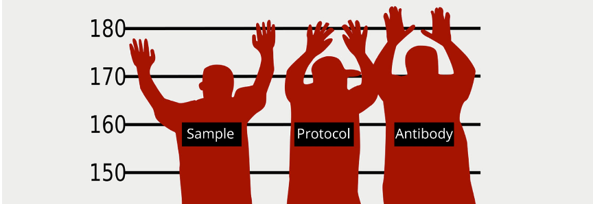
An immunohistochemistry experiment is a combination of 3 major elements: sample, application and experimental protocol. An excellent IHC staining is achieved when all the required elements, qualities and characteristics are matched perfectly. A combination as good as it is possible be.
So, if you are looking for suspects to blame for the artifact in your IHC experiment, keep in mind the 3 usual suspects:
- Sample (tissue or cell)
- Antibodies (primary and secondary)
- IHC protocol
For advice on how to choose an antibody, read our previous post.
Look carefully at the table below: these tips will help you to identify the possible culprit.
|
Evidence: Lack of Staining |
Evidence: High Background | Evidence: Non Specific Binding | |
| Suspect: Sample | Sample does not express the antigen. | Presence of Fc receptors that bind the Fc region of antibodies. | |
|
Sections are too tick, preventing penetration of antibodies. |
Sample is damaged or necrotic. | Sample dried out. | |
| Sample is insufficiently fixated or over fixated. | |||
| Sample dried out. | |||
| Suspect: Antibody | Primary antibody does not work in your application. | Non specific binding between antibody’s Fc portion and endogenous Fc receptors. | |
| Primary antibody cannot detect target protein in native conformation (not an issue for IHC-using FFPE). | Antibodies not diluted enough. | ||
| Improper antibodies storage. | |||
| Incompatible antibodies. | |||
| Suspect: IHC Protocol | Lack of, or inappropriate antigen retrieval. | Reagent incompatibility or insufficient washing. | |
| Excessive wash buffer. | Improper fixative, prolonged fixation time and interval before fixation. | ||
| Excessive blocking solution. |
Insufficient rinsing time. |
Endogenous enzyme activities are not blocked. |
|
| Inadequate chromogenic detection. | Inappropriate detection method. | Wrong temperature or pH during antigen retrieval. | |
|
Protracted time of chromogen application. |
|
||
In Need of Troubleshooting Advice?
OK, you have an idea now what to blame for the artifacts in your IHC staining.
That sounds good enough, but when it comes time to do, to solve those artifacts, it it's hard to know where to start. For help on how to proceed, keep reading the second part in our next blog: Artifacts in IHC: the Usual Suspects - Part II.
Recommended Reading
Buchwalow I. et al. Sci Rep. (2011) 1: 28, Non-specific binding of antibodies in immunohistochemistry: fallacies and facts.
De Matos L.L., et al. Biomarker Insights (2010):5 9–20, Immunohistochemistry as an Important Tool in Biomarkers Detection and clinical practice.
Ramos-Vara J.A. and Miller M. A. Veterinary Pathology (2014) 51(1) 42-87, When tissue antigens and antibodies get along: revisiting the technical aspects of immunohistochemistry: the red, brown, and blue technique.
Ward J.M. and Rehg J.E. Veterinary Pathology, (2013) 51 (1) 88-101, Rodent Immunohistochemistry: Pitfalls and Troubleshooting.
Written by Dr. Laura Pozzi
Dr. Laura Pozzi is a scientific writer at Atlas Antibodies. She holds a Ph.D. in Life and Biomolecular Science from the Open University of London in collaboration with the Mario Negri Institute for Pharmacological Research in Milan. Laura has worked as a researcher at Karolinska Institutet in Sweden and more recently as associated editor. She has a long track record of scientific publications as a first author and as coauthor. Her research focus on neuroscience with a broad experience in antibodies validation and immunohistochemistry techniques.


