
CD79a Rabbit pAb, Unconjugated
Catalog Number:
ABB-A0331
- Images (7)


| Article Name: | CD79a Rabbit pAb, Unconjugated |
| Biozol Catalog Number: | ABB-A0331 |
| Supplier Catalog Number: | A0331 |
| Alternative Catalog Number: | ABB-A0331-100UL,ABB-A0331-20UL,ABB-A0331-500UL,ABB-A0331-1000UL |
| Manufacturer: | ABclonal |
| Host: | Rabbit |
| Category: | Antikörper |
| Application: | ELISA, IF, IHC-P, WB |
| Species Reactivity: | Human |
| Immunogen: | Recombinant protein (or fragment).This information is considered to be commercially sensitive. |
| Conjugation: | Unconjugated |
| Alternative Names: | IGA, MB1, MB-1, IGAlpha, CD79a |
| The B lymphocyte antigen receptor is a multimeric complex that includes the antigen-specific component, surface immunoglobulin (Ig). Surface Ig non-covalently associates with two other proteins, Ig-alpha and Ig-beta, which are necessary for expression and function of the B-cell antigen receptor. This gene encodes the Ig-alpha protein of the B-cell antigen component. Alternatively spliced transcript variants encoding different isoforms have been described. |
| Application Dilute: | WB,1:500 - 1:1000|IF-P,1:50 - 1:200|IHC-P,1:50 - 1:200|ELISA,Recommended starting concentration is 1 µg/mL. Please optimize the concentration based on your specific assay requirements. |
| Application Notes: | Cross-Reactivity: Human,Mouse,Rat, ResearchArea: Cancer,Tumor immunology,Immunology Inflammation,CDs,B Cell Receptor Signaling Pathway,Stem Cells,Hematopoietic Progenitors. |
|
|
Western blot analysis of various lysates using CD79a Rabbit pAb (A0331) at 1:1000 dilution. Secondary antibody: HRP-conjugated Goat anti-Rabbit IgG (H+L) (AS014) at 1:10000 dilution. Lysates/proteins: 25µg per lane. Blocking buffer: 3% nonfat dry milk in TBST. Detection: ECL Basic Kit (RM00020). Exposure time: 10s. |
|
|
Immunohistochemistry analysis of paraffin-embedded Rat spleen using CD79a Rabbit pAb (A0331) at dilution of 1:100 (40x lens). High pressure antigen retrieval performed with 0.01M Citrate buffer (pH 6.0) prior to IHC staining. |
|
|
Western blot analysis of lysates from Raji cells, using CD79a Rabbit pAb (A0331) at 1:1000 dilution. Secondary antibody: HRP-conjugated Goat anti-Rabbit IgG (H+L) (AS014) at 1:10000 dilution. Lysates/proteins: 25µg per lane. Blocking buffer: 3% nonfat dry milk in TBST. Detection: ECL Basic Kit (RM00020). Exposure time: 1s. |
|
|
Immunohistochemistry analysis of paraffin-embedded Human spleen using CD79a Rabbit pAb (A0331) at dilution of 1:100 (40x lens). High pressure antigen retrieval performed with 0.01M Citrate buffer (pH 6.0) prior to IHC staining. |
|
|
Immunohistochemistry analysis of paraffin-embedded Mouse spleen using CD79a Rabbit pAb (A0331) at dilution of 1:100 (40x lens). High pressure antigen retrieval performed with 0.01M Citrate buffer (pH 6.0) prior to IHC staining. |
|
|
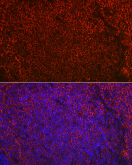
Immunofluorescence analysis of paraffin-embedded rat spleen using CD79a Rabbit pAb (A0331) at dilution of 1:50 (40x lens). Secondary antibody: Cy3-conjugated Goat anti-Rabbit IgG (H+L) (AS007) at 1:500 dilution. Blue: DAPI for nuclear staining. |
|
|
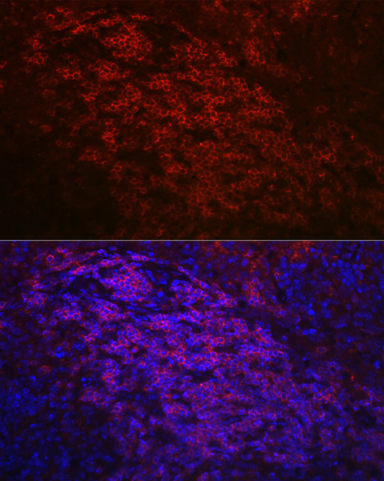
Immunofluorescence analysis of paraffin-embedded mouse spleen using CD79a Rabbit pAb (A0331) at dilution of 1:50 (40x lens). Secondary antibody: Cy3-conjugated Goat anti-Rabbit IgG (H+L) (AS007) at 1:500 dilution. Blue: DAPI for nuclear staining. |
Product Guarantee and Expert Support
