
YAP1 Rabbit pAb, Unconjugated, Polyclonal
Catalog Number:
ABB-A1002
- Images (9)


| Article Name: | YAP1 Rabbit pAb, Unconjugated, Polyclonal |
| Biozol Catalog Number: | ABB-A1002 |
| Supplier Catalog Number: | A1002 |
| Alternative Catalog Number: | ABB-A1002-100UL,ABB-A1002-20UL,ABB-A1002-1000UL,ABB-A1002-500UL |
| Manufacturer: | ABclonal |
| Host: | Rabbit |
| Category: | Antikörper |
| Application: | ELISA, IF, IHC-P, IP, WB |
| Species Reactivity: | Human |
| Immunogen: | Recombinant protein (or fragment).This information is considered to be commercially sensitive. |
| Conjugation: | Unconjugated |
| Alternative Names: | YAP, YKI, COB1, YAP2, YAP-1, YAP65, YAP1 |
| This gene encodes a downstream nuclear effector of the Hippo signaling pathway which is involved in development, growth, repair, and homeostasis. This gene is known to play a role in the development and progression of multiple cancers as a transcriptional regulator of this signaling pathway and may function as a potential target for cancer treatment. Alternative splicing results in multiple transcript variants encoding different isoforms. |
| Clonality: | Polyclonal |
| Molecular Weight: | 54kDa |
| NCBI: | 10413 |
| UniProt: | P46937 |
| Purity: | Affinity purification |
| Sequence: | PTAQHLRQSSFEIPDDVPLPAGWEMAKTSSGQRYFLNHIDQTTTWQDPRKAMLSQMNVTAPTSPPVQQNMMNSASGPLPDGWEQAMTQDGEIYYINHKNKTTSWLDPRLDPRFAMNQRISQSAPVKQPPPLAPQSPQGGVMGGSNSNQQQQMRLQQLQMEKERLRLKQQELLRQAMRNINPSTANSPKCQELALRSQLPTLEQDGGTQNPVSSPGMSQELRTMTTNSSDPFLNSGTYHSRDESTDSGLSMSSYSV |
| Target: | YAP1 |
| Antibody Type: | Primary Antibody |
| Application Dilute: | WB,1:2000 - 1:20000|IHC-P,1:500 - 1:1000|IF/ICC,1:50 - 1:200|IP,0.5µg-4µg antibody for 200µg-400µg extracts of whole cells|ELISA,Recommended starting concentration is 1 µg/mL. Please optimize the concentration based on your specific assay requirements. |
| Application Notes: | Cross-Reactivity: Human,Mouse. ResearchArea: Epigenetics Nuclear Signaling,Transcription Factors,Protein phosphorylation,Cancer,Signal Transduction,Kinase,Tyrosine kinases,Cell Biology Developmental Biology. Shipping: Ice Bag |
|
|
Immunohistochemistry analysis of paraffin-embedded Human colon (low expression samples) using YAP1 Rabbit pAb (A1002) at dilution of 1:1000 (40x lens). High pressure antigen retrieval performed with 0.01M Citrate buffer (pH 6.0) prior to IHC staining. |
|
|
Immunohistochemistry analysis of paraffin-embedded Human colon carcinoma using YAP1 Rabbit pAb (A1002) at dilution of 1:1000 (40x lens). High pressure antigen retrieval performed with 0.01M Citrate buffer (pH 6.0) prior to IHC staining. |
|
|
Western blot analysis of lysates from wild type (WT) and YAP1 knockout (KO)HeLa cellsusingYAP1 Rabbit pAb (A1002) at1:2000 dilution. Secondary antibody: HRP-conjugated Goat anti-Rabbit IgG (H+L) (AS014) at 1:10000 dilution. Lysates/proteins: 25µg per lane. Blocking buffer: 3% nonfat dry milk in TBST. Detection: ECL Basic Kit (RM00020). Exposure time:30s. |
|
|
Immunohistochemistry analysis of paraffin-embedded Human liver cancer using YAP1 Rabbit pAb (A1002) at dilution of 1:1000 (40x lens). High pressure antigen retrieval performed with 0.01M Citrate buffer (pH 6.0) prior to IHC staining. |
|
|
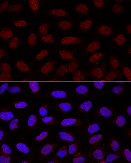
Confocal immunofluorescence analysis of U2OS cells using YAP1 Rabbit pAb (A1002) at dilution of 1:200. Blue: DAPI for nuclear staining. |
|
|
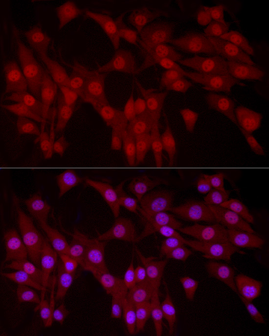
Immunofluorescence analysis of NIH/3T3 cells using YAP1 Rabbit pAb (A1002) at dilution of 1:100 (40x lens). Secondary antibody: Cy3-conjugated Goat anti-Rabbit IgG (H+L) (AS007) at 1:500 dilution. Blue: DAPI for nuclear staining. |
|
|
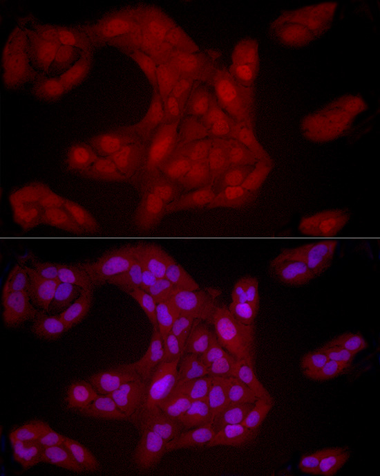
Immunofluorescence analysis of U2OS cells using YAP1 Rabbit pAb (A1002) at dilution of 1:100 (40x lens). Secondary antibody: Cy3-conjugated Goat anti-Rabbit IgG (H+L) (AS007) at 1:500 dilution. Blue: DAPI for nuclear staining. |
|
|
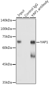
Immunoprecipitation analysis of 300 µg extracts of HeLa cells using 3 µg YAP1 antibody (A1002). Western blot was performed from the immunoprecipitate using YAP1 antibody (A1002) at a dilution of 1:1000. |
|
|
Immunoprecipitation analysis of 300 µg extracts of HeLa cells using 3 µg YAP1 antibody (A1002). Western blot was performed from the immunoprecipitate using YAP1 (A1002) at a dilution of 1:1000. |
Product Guarantee and Expert Support
