
[KD Validated] CDX2 Rabbit mAb, Unconjugated, Monoclonal
Catalog Number:
ABB-A19030
- Images (9)


| Article Name: | [KD Validated] CDX2 Rabbit mAb, Unconjugated, Monoclonal |
| Biozol Catalog Number: | ABB-A19030 |
| Supplier Catalog Number: | A19030 |
| Alternative Catalog Number: | ABB-A19030-100UL,ABB-A19030-20UL,ABB-A19030-1000UL,ABB-A19030-500UL |
| Manufacturer: | ABclonal |
| Host: | Rabbit |
| Category: | Antikörper |
| Application: | ELISA, IF, IHC-P, IP, WB |
| Species Reactivity: | Human |
| Immunogen: | Synthetic peptide. This information is considered to be commercially sensitive. |
| Conjugation: | Unconjugated |
| Alternative Names: | CDX3, CDX-3, CDX2/AS, X2 |
| This gene is a member of the caudal-related homeobox transcription factor gene family. The encoded protein is a major regulator of intestine-specific genes involved in cell growth an differentiation. This protein also plays a role in early embryonic development of the intestinal tract. Aberrant expression of this gene is associated with intestinal inflammation and tumorigenesis. |
| Application Dilute: | WB,1:500 - 1:2000|IP,0.5µg-4µg antibody for 200µg-600µg extracts of whole cells|IF-P,1:100 - 1:1000|IHC-P,1:3000 - 1:12000|ELISA,Recommended starting concentration is 1 µg/mL. Please optimize the concentration based on your specific assay requirements. |
| Application Notes: | Cross-Reactivity: Human,Mouse,Rat. ResearchArea: Epigenetics Nuclear Signaling,Transcription Factors,Cell Biology Developmental Biology,Stem Cells. Shipping: Ice Bag |
|
|
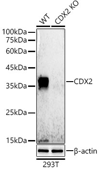
Western blot analysis of lysates from wild type(WT) and CDX2 knockdown (KD) 293T cells, using [KD Validated] CDX2 Rabbit mAb (A19030) at 1:500 dilution. Secondary antibody: HRP-conjugated Goat anti-Rabbit IgG (H+L) (AS014) at 1:10000 dilution. Lysates/proteins: 25µg per lane. Blocking buffer: 3% nonfat dry milk in TBST. Detection: ECL Basic Kit (RM00020). Exposure time: 30s. |
|
|
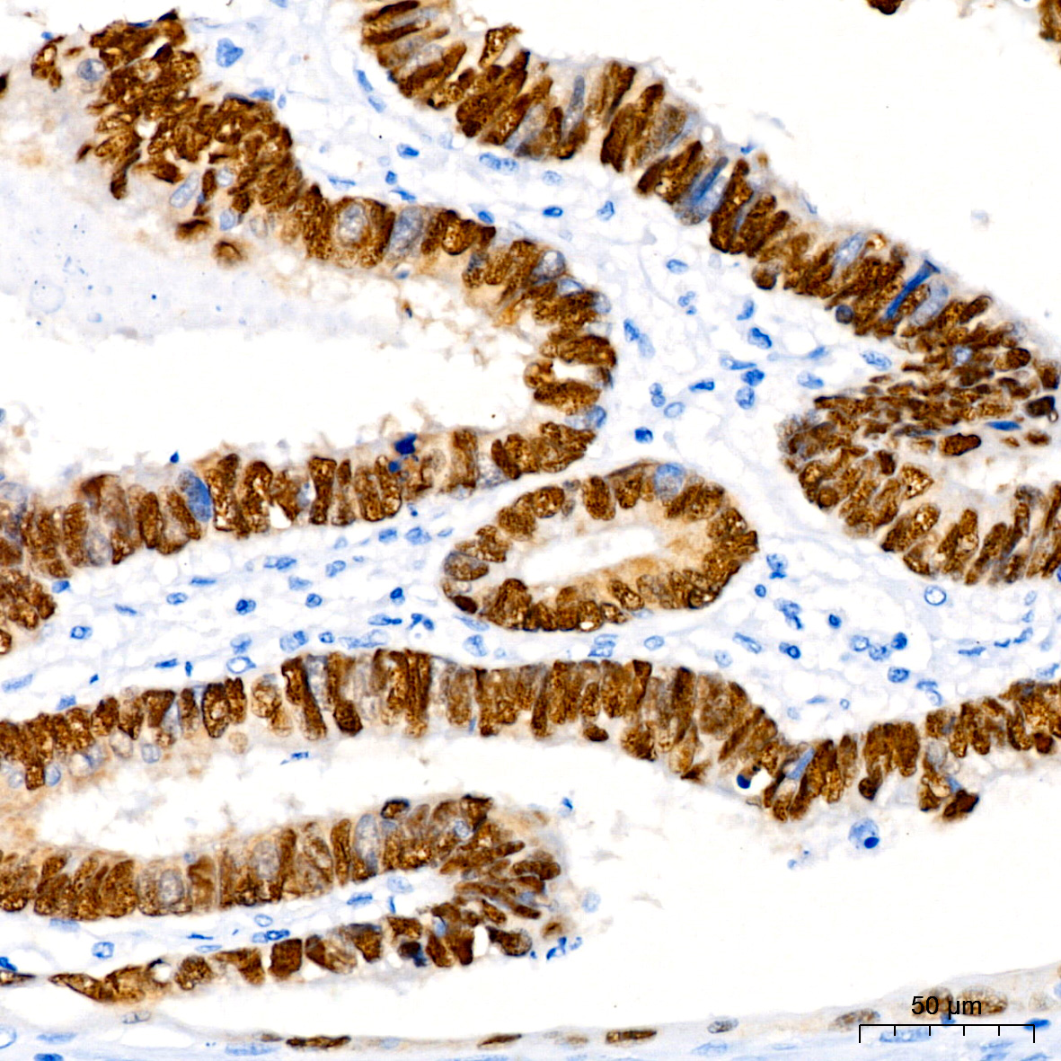
Immunohistochemistry analysis of paraffin-embedded Human colon tissue using [KD Validated] CDX2 Rabbit mAb (A19030) at a dilution of 1:4000 (40x lens). High pressure antigen retrieval performed with 0.01M Tris-EDTA Buffer (pH 9.0) prior to IHC staining. |
|
|
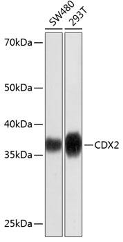
Western blot analysis of various lysates using [KD Validated] CDX2 Rabbit mAb (A19030) at 1:1000 dilution. Secondary antibody: HRP-conjugated Goat anti-Rabbit IgG (H+L) (AS014) at 1:10000 dilution. Lysates/proteins: 25µg per lane. Blocking buffer: 3% nonfat dry milk in TBST. Detection: ECL Basic Kit (RM00020). Exposure time: 3min. |
|
|
Immunohistochemistry analysis of paraffin-embedded Human pancreas tissue using [KD Validated] CDX2 Rabbit mAb (A19030) at a dilution of 1:4000 (40x lens). High pressure antigen retrieval performed with 0.01M Tris-EDTA Buffer (pH 9.0) prior to IHC staining. |
|
|
Immunohistochemistry analysis of paraffin-embedded Mouse colon tissue using [KD Validated] CDX2 Rabbit mAb (A19030) at a dilution of 1:4000 (40x lens). High pressure antigen retrieval performed with 0.01M Tris-EDTA Buffer (pH 9.0) prior to IHC staining. |
|
|
Immunohistochemistry analysis of paraffin-embedded Rat colon tissue using [KD Validated] CDX2 Rabbit mAb (A19030) at a dilution of 1:4000 (40x lens). High pressure antigen retrieval performed with 0.01M Tris-EDTA Buffer (pH 9.0) prior to IHC staining. |
|
|
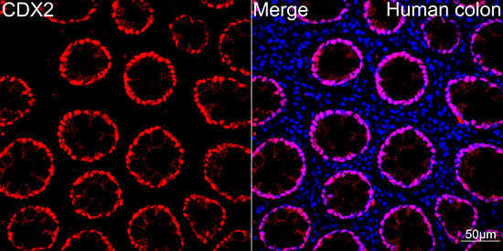
Confocal imaging of Human colon using [KD Validated] CDX2 Rabbit mAb (A19030,dilution 1:100)(Red). DAPI was used for nuclear staining (blue). Objective: 40x. |
|
|
Immunoprecipitation analysis of 600 µg extracts of 293T cells using 3 µg [KD Validated] CDX2 Rabbit mAb (A19030). Western blot was performed from the immunoprecipitate using CDX2 antibody (A19030) at a dilution of 1:500. |
|
|
Immunoprecipitation analysis of 600 µg extracts of 293T cells using 3 µg [KD Validated] CDX2 Rabbit mAb (A19030). Western blot was performed from the immunoprecipitate using CDX2 antibody (A19030) at a dilution of 1:500. |
Product Guarantee and Expert Support
