
[KD Validated] METTL3 Rabbit mAb, Unconjugated, Monoclonal
Catalog Number:
ABB-A19079
- Images (9)

| Article Name: | [KD Validated] METTL3 Rabbit mAb, Unconjugated, Monoclonal |
| Biozol Catalog Number: | ABB-A19079 |
| Supplier Catalog Number: | A19079 |
| Alternative Catalog Number: | ABB-A19079-100UL,ABB-A19079-20UL,ABB-A19079-1000UL,ABB-A19079-500UL |
| Manufacturer: | ABclonal |
| Host: | Rabbit |
| Category: | Antikörper |
| Application: | ELISA, IF, IHC-P, IP, WB |
| Species Reactivity: | Human |
| Immunogen: | Recombinant protein (or fragment).This information is considered to be commercially sensitive. |
| Conjugation: | Unconjugated |
| Alternative Names: | M6A, IME4, Spo8, MT-A70, hMETTL3, METTL3 |
| This gene encodes the 70 kDa subunit of MT-A which is part of N6-adenosine-methyltransferase. This enzyme is involved in the posttranscriptional methylation of internal adenosine residues in eukaryotic mRNAs, forming N6-methyladenosine. |
| Clonality: | Monoclonal |
| Clone Designation: | [ARC0487] |
| Molecular Weight: | 64kDa |
| NCBI: | 56339 |
| UniProt: | Q86U44 |
| Purity: | Affinity purification |
| Sequence: | SDTWSSIQAHKKQLDSLRERLQRRRKQDSGHLDLRNPEAALSPTFRSDSPVPTAPTSGGPKPSTASAVPELATDPELEKKLLHHLSDLALTLPTDAVSICLAISTPDAPATQDGVESLLQKFAAQELIEVKRGLLQDDAHPTLVTYADHSKLSAMMGAVAEKKGPGEVAGTVTGQKRRAEQDSTTVAAFASSLVSGLNSSASEPAKEPAKKSRKHAASDVDLEIESLLNQQSTKEQQSKKVSQEILELL |
| Target: | METTL3 |
| Antibody Type: | Primary Antibody |
| Application Dilute: | WB,1:1000 - 1:2000|IP,0.5µg-4µg antibody for 200µg-400µg extracts of whole cells|IF/ICC,1:100 - 1:400|IHC-P,1:200 - 1:800|ELISA,Recommended starting concentration is 1 µg/mL. Please optimize the concentration based on your specific assay requirements. |
| Application Notes: | Cross-Reactivity: Human,Mouse,Rat. ResearchArea: Epigenetics Nuclear Signaling. Shipping: Ice Bag |
|
|
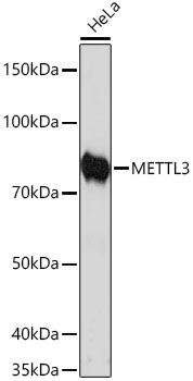
Western blot analysis of lysates from HeLa cells using [KD Validated] METTL3 Rabbit mAb (A19079) at 1:1000 dilution incubated overnight at 4°C. Secondary antibody: HRP-conjugated Goat anti-Rabbit IgG (H+L) (AS014) at 1:10000 dilution. Lysates/proteins: 25 µg per lane. Blocking buffer: 3% nonfat dry milk in TBST. Detection: ECL Basic Kit (RM00020). Exposure time: 20s. |
|
|
Western blot analysis of lysates from Rat brain using [KD Validated] METTL3 Rabbit mAb (A19079) at 1:1000 dilution incubated overnight at 4°C. Secondary antibody: HRP-conjugated Goat anti-Rabbit IgG (H+L) (AS014) at 1:10000 dilution. Lysates/proteins: 25 µg per lane. Blocking buffer: 3% nonfat dry milk in TBST. Detection: ECL Basic Kit (RM00020). Exposure time: 30s. |
|
|
Western blot analysis of lysates from wild type (WT) and METTL3 knockdown (KD) 293T cells using METTL3 Rabbit mAb (A19079) at 1:1000 dilution incubated overnight at 4°C. Secondary antibody: HRP-conjugated Goat anti-Rabbit IgG (H+L) (AS014) at 1:10000 dilution. Lysates/proteins: 25 µg per lane. Blocking buffer: 3% nonfat dry milk in TBST. Detection: ECL Basic Kit (RM00020). Exposure time: 20s. |
|
|
Western blot analysis of lysates from Mouse brain using [KD Validated] METTL3 Rabbit mAb (A19079) at 1:1000 dilution incubated overnight at 4°C. Secondary antibody: HRP-conjugated Goat anti-Rabbit IgG (H+L) (AS014) at 1:10000 dilution. Lysates/proteins: 25 µg per lane. Blocking buffer: 3% nonfat dry milk in TBST. Detection: ECL Basic Kit (RM00020). Exposure time: 60s. |
|
|
Immunohistochemistry analysis of paraffin-embedded Mouse testis tissue using [KD Validated] METTL3 Rabbit mAb (A19079) at a dilution of 1:200 (40x lens). High pressure antigen retrieval performed with 0.01M Tris-EDTA Buffer (pH 9.0) prior to IHC staining. |
|
|
Immunohistochemistry analysis of paraffin-embedded Human tonsil tissue using [KD Validated] METTL3 Rabbit mAb (A19079) at a dilution of 1:200 (40x lens). High pressure antigen retrieval performed with 0.01M Tris-EDTA Buffer (pH 9.0) prior to IHC staining. |
|
|
Immunohistochemistry analysis of paraffin-embedded Rat spleen tissue using [KD Validated] METTL3 Rabbit mAb (A19079) at a dilution of 1:200 (40x lens). High pressure antigen retrieval performed with 0.01M Tris-EDTA Buffer (pH 9.0) prior to IHC staining. |
|
|
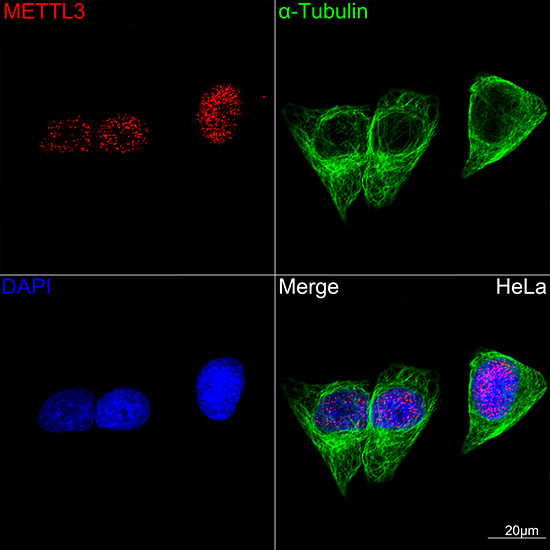
Confocal imaging of HeLa cells using [KD Validated] METTL3 Rabbit mAb (A19079,dilution 1:100)(Red) followed by a further incubation with Cy3-conjugated Goat Anti-Rabbit IgG (H+L) (AS007, dilution 1:500) (Red). The cells were counterstained with alpha-Tubulin mAb (AC012, dilution 1:400) followed by incubation with ABflo 488-conjugated Goat Anti-Mouse IgG (H+L) (AS076, dilution 1:500) (Green). DAPI was used for nuclear staining (blue). Objective: 100x. |
|
|
Immunoprecipitation of METTL3 from 200 µg extracts of 293F cells was performed using 0.5 µg of [KD Validated] METTL3 Rabbit mAb (A19079). Rabbit IgG isotype control(AC042) was used to precipitate the Control IgG sample. IP samples were eluted with 1x reducing Laemmli Buffer. The Input lane represents 10% of the total input. Western blot analysis of immunoprecipitates was conducted using [KD Validated] METTL3 Rabbit mAb (A19079) at a dilution of 1:500. |
Product Guarantee and Expert Support
