
[KO Validated] N-Cadherin Rabbit mAb, Unconjugated, Monoclonal
Catalog Number:
ABB-A19083
- Images (9)

| Article Name: | [KO Validated] N-Cadherin Rabbit mAb, Unconjugated, Monoclonal |
| Biozol Catalog Number: | ABB-A19083 |
| Supplier Catalog Number: | A19083 |
| Alternative Catalog Number: | ABB-A19083-20UL,ABB-A19083-100UL,ABB-A19083-500UL,ABB-A19083-1000UL |
| Manufacturer: | ABclonal |
| Host: | Rabbit |
| Category: | Antikörper |
| Application: | ELISA, IF, IHC-P, WB |
| Species Reactivity: | Human |
| Immunogen: | Synthetic peptide. This information is considered to be commercially sensitive. |
| Conjugation: | Unconjugated |
| Alternative Names: | CDHN, NCAD, ACOGS, ADHD8, CD325, ARVD14, CDw325, in |
| This gene encodes a classical cadherin and member of the cadherin superfamily. Alternative splicing results in multiple transcript variants, at least one of which encodes a preproprotein is proteolytically processed to generate a calcium-dependent cell adhesion molecule and glycoprotein. This protein plays a role in the establishment of left-right asymmetry, development of the nervous system and the formation of cartilage and bone. |
| Application Dilute: | WB,1:1000 - 1:2000|IF-P,1:50 - 1:200|IHC-P,1:1000 - 1:4000|ELISA,Recommended starting concentration is 1 µg/mL. Please optimize the concentration based on your specific assay requirements. |
| Application Notes: | Cross-Reactivity: Human,Mouse,Rat. ResearchArea: Cancer,Invasion and Metastasis,Signal Transduction,Cell Biology Developmental Biology,Cell Cycle,Centrosome,Cell Adhesion,Cadherins,Tight Junctions,Cytoskeleton,Wnt -Catenin Signaling Pathway,Immunology Inflammation,CDs,Stem Cells,Hematopoietic Progenitors. Shipping: Ice Bag |
|
|
Western blot analysis of lysates from C2C12 cells using [KO Validated] N-Cadherin Rabbit mAb (A19083) at 1:1000 dilution incubated overnight at 4°C. Secondary antibody: HRP-conjugated Goat anti-Rabbit IgG (H+L) (AS014) at 1:10000 dilution. Lysates/proteins: 25 µg per lane. Blocking buffer: 3% nonfat dry milk in TBST. Detection: ECL Basic Kit (RM00020). Exposure time: 60s. |
|
|
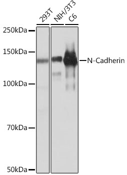
Western blot analysis of lysates from C6 cells using [KO Validated] N-Cadherin Rabbit mAb (A19083) at 1:1000 dilution incubated overnight at 4°C. Secondary antibody: HRP-conjugated Goat anti-Rabbit IgG (H+L) (AS014) at 1:10000 dilution. Lysates/proteins: 25 µg per lane. Blocking buffer: 3% nonfat dry milk in TBST. Detection: ECL Basic Kit (RM00020). Exposure time: 10s. |
|
|
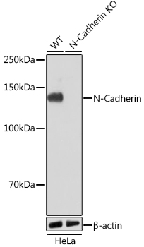
Western blot analysis of lysates from wild type (WT) and N-Cadherin knockout (KO) HeLa cells using [KO Validated] N-Cadherin Rabbit mAb (A19083) at 1:1000 dilution incubated overnight at 4°C. Secondary antibody: HRP-conjugated Goat anti-Rabbit IgG (H+L) (AS014) at 1:10000 dilution. Lysates/proteins: 25µg per lane. Blocking buffer: 3% nonfat dry milk in TBST. Detection: ECL Basic Kit (RM00020). Exposure time: 1min. |
|
|
Immunohistochemistry analysis of paraffin-embedded Human liver tissue using [KO Validated] N-Cadherin Rabbit mAb (A19083) at a dilution of 1:2000 (40x lens). High pressure antigen retrieval performed with 0.01M Tris-EDTA Buffer (pH 9.0) prior to IHC staining. |
|
|
Immunohistochemistry analysis of paraffin-embedded Mouse brain tissue using [KO Validated] N-Cadherin Rabbit mAb (A19083) at a dilution of 1:2000 (40x lens). High pressure antigen retrieval performed with 0.01M Tris-EDTA Buffer (pH 9.0) prior to IHC staining. |
|
|
Immunohistochemistry analysis of paraffin-embedded Mouse colon tissue using [KO Validated] N-Cadherin Rabbit mAb (A19083) at a dilution of 1:2000 (40x lens). High pressure antigen retrieval performed with 0.01M Tris-EDTA Buffer (pH 9.0) prior to IHC staining. |
|
|
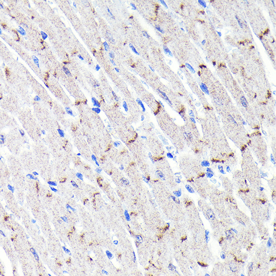
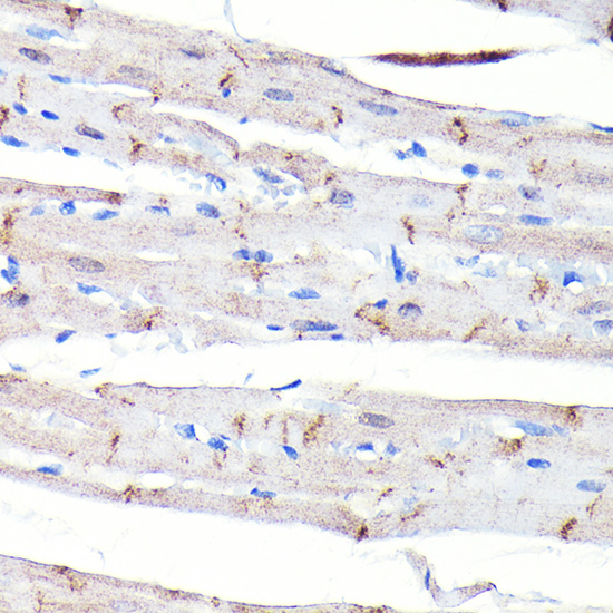
Immunohistochemistry analysis of paraffin-embedded Rat brain tissue using [KO Validated] N-Cadherin Rabbit mAb (A19083) at a dilution of 1:2000 (40x lens). High pressure antigen retrieval performed with 0.01M Tris-EDTA Buffer (pH 9.0) prior to IHC staining. |
|
|
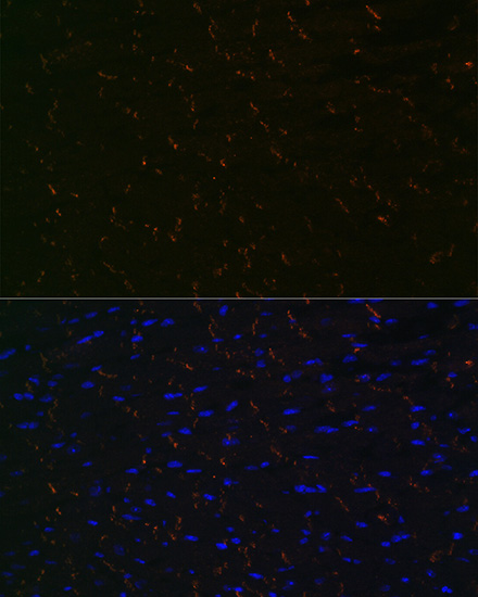
Confocal imaging of paraffin-embedded rat heart using [KO Validated] N-Cadherin Rabbit mAb (A19083, dilution 1:200) followed by a further incubation with Cy3 Goat Anti-Rabbit IgG (H+L) (AS007, dilution 1:500) (Red). DAPI was used for nuclear staining (Blue). Objective: 40x. High pressure antigen retrieval performed with 0.01M Citrate Buffer(pH 6.0) prior to IF staining. |
|
|
Confocal imaging of paraffin-embedded Mouse heart using [KO Validated] N-Cadherin Rabbit mAb (A19083, dilution 1:200) followed by a further incubation with Cy3 Goat Anti-Rabbit IgG (H+L) (AS007, dilution 1:500) (Red). DAPI was used for nuclear staining (Blue). Objective: 40x. High pressure antigen retrieval performed with 0.01M Citrate Buffer(pH 6.0) prior to IF staining. |
Product Guarantee and Expert Support
