
[KD Validated] beta 2 Microglobulin Rabbit mAb, Unconjugated, Monoclonal
Catalog Number:
ABB-A23430
- Images (9)


| Article Name: | [KD Validated] beta 2 Microglobulin Rabbit mAb, Unconjugated, Monoclonal |
| Biozol Catalog Number: | ABB-A23430 |
| Supplier Catalog Number: | A23430 |
| Alternative Catalog Number: | ABB-A23430-100UL,ABB-A23430-500UL,ABB-A23430-20UL,ABB-A23430-1000UL |
| Manufacturer: | ABclonal |
| Host: | Rabbit |
| Category: | Antikörper |
| Application: | ELISA, FC, IF, IHC-P, WB |
| Species Reactivity: | Human |
| Immunogen: | Recombinant protein (or fragment).This information is considered to be commercially sensitive. |
| Conjugation: | Unconjugated |
| Alternative Names: | B2M, IMD43, beta-2-microglobulin, [KD Validated] beta 2 Microglobulin |
| This gene encodes a serum protein found in association with the major histocompatibility complex (MHC) class I heavy chain on the surface of nearly all nucleated cells. The protein has a predominantly beta-pleated sheet structure that can form amyloid fibrils in some pathological conditions. The encoded antimicrobial protein displays antibacterial activity in amniotic fluid. A mutation in this gene has been shown to result in hypercatabolic hypoproteinemia. |
| Application Dilute: | WB,1:1000 - 1:4000|IHC-P,1:1000 - 1:4000|IF/ICC,1:50 - 1:200|FC,1:500 - 1:1000|ELISA,Recommended starting concentration is 1 µg/mL. Please optimize the concentration based on your specific assay requirements. |
| Application Notes: | Cross-Reactivity: Human. ResearchArea: Cancer,Tumor biomarkers,Cardiovascular,Blood,Blood Cell Antigens,Serum Proteins. Shipping: Ice Bag |
|
|
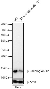
Western blot analysis of lysates from wild type(WT) and beta2-microglobulin knockdown (KD) HeLa cells, using [KD Validated] beta 2 Microglobulin Rabbit mAb (A23430) at 1:1000 dilution. Secondary antibody: HRP-conjugated Goat anti-Rabbit IgG (H+L) (AS014) at 1:10000 dilution. Lysates/proteins: 25µg per lane. Blocking buffer: 3% nonfat dry milk in TBST. Detection: ECL Basic Kit (RM00020). Exposure time: 30s. |
|
|
Immunohistochemistry analysis of paraffin-embedded Human esophagus tissue using [KD Validated] beta 2 Microglobulin Rabbit mAb (A23430) at a dilution of 1:2000 (40x lens). High pressure antigen retrieval performed with 0.01M Tris-EDTA Buffer (pH 9.0) prior to IHC staining. |
|
|
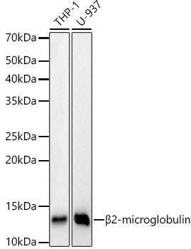
Western blot analysis of various lysates, using [KD Validated] beta 2 Microglobulin Rabbit mAb (A23430) at 1:1000 dilution. Secondary antibody: HRP-conjugated Goat anti-Rabbit IgG (H+L) (AS014) at 1:10000 dilution. Lysates/proteins: 25µg per lane. Blocking buffer: 3% nonfat dry milk in TBST. Detection: ECL Basic Kit (RM00020). Exposure time: 30s. |
|
|
Immunohistochemistry analysis of paraffin-embedded Human colon carcinoma tissue using [KD Validated] beta 2 Microglobulin Rabbit mAb (A23430) at a dilution of 1:2000 (40x lens). High pressure antigen retrieval performed with 0.01M Tris-EDTA Buffer (pH 9.0) prior to IHC staining. |
|
|
Immunohistochemistry analysis of paraffin-embedded Human liver cancer tissue using [KD Validated] beta 2 Microglobulin Rabbit mAb (A23430) at a dilution of 1:2000 (40x lens). High pressure antigen retrieval performed with 0.01M Tris-EDTA Buffer (pH 9.0) prior to IHC staining. |
|
|
Immunohistochemistry analysis of paraffin-embedded Human tonsil tissue using [KD Validated] beta 2 Microglobulin Rabbit mAb (A23430) at a dilution of 1:2000 (40x lens). High pressure antigen retrieval performed with 0.01M Tris-EDTA Buffer (pH 9.0) prior to IHC staining. |
|
|
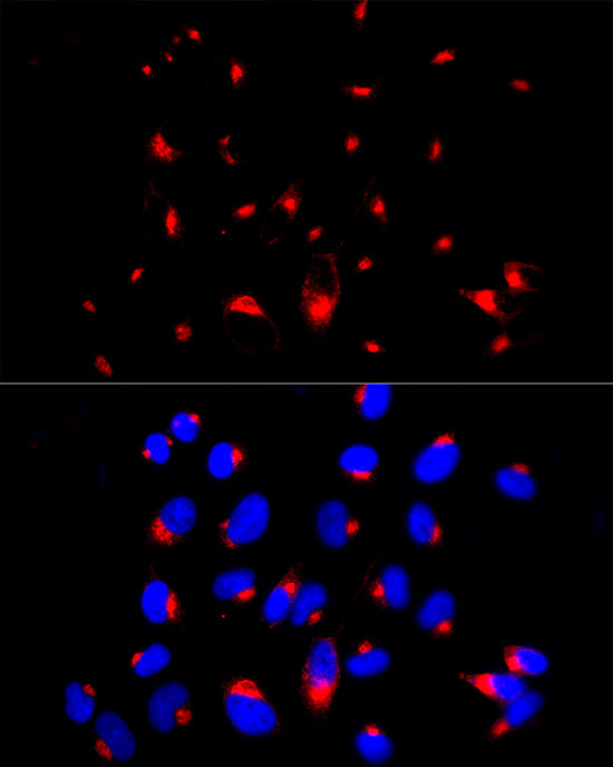
Immunofluorescence analysis of HeLa cells using [KD Validated] beta 2 Microglobulin Rabbit mAb (A23430) at dilution of 1:100 (40x lens). Secondary antibody: Cy3-conjugated Goat anti-Rabbit IgG (H+L) (AS007) at 1:500 dilution. Blue: DAPI for nuclear staining. |
|
|
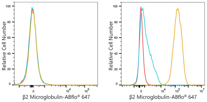
Flow cytometry:1X10 6 Daudi cells(negative control,left) and Hela cells (right) were surface-stained with [KD Validated] beta Microglobulin Rabbit mAb(A23430,2 µg/mL,orange line) or ABflo 647 Rabbit IgG isotype control (A22070,2 µg/mL,blue line).Non-fluorescently stained cells was used as blank control (red line). |
|
|
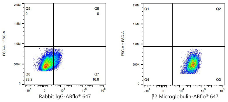
Flow cytometry:1X10 6 HeLa cells were surface-stained with ABflo 647 Rabbit IgG isotype control (A22070,2 µg/mL,left) or beta2 Microglobulin Rabbit mAb(A23430,2 µg/mL,right). |
Product Guarantee and Expert Support
