
CD3 Intracellular domain Rabbit Polyclonal Antibody, Unconjugated
Catalog Number:
BYT-ORB1294242
- Images (8)


| Article Name: | CD3 Intracellular domain Rabbit Polyclonal Antibody, Unconjugated |
| Biozol Catalog Number: | BYT-ORB1294242 |
| Supplier Catalog Number: | orb1294242 |
| Alternative Catalog Number: | BYT-ORB1294242-25,BYT-ORB1294242-100 |
| Manufacturer: | Biorbyt |
| Host: | Rabbit |
| Category: | Antikörper |
| Application: | IF, IHC, WB |
| Species Reactivity: | Human, Mouse, Rat |
| Immunogen: | Synthetic peptide / Intracellular domain corresponding to Human/mouse CD3 |
| Conjugation: | Unconjugated |
| Rabbit polyclonal antibody to CD3 (Intracellular |
| Clonality: | Polyclonal |
| Buffer: | 100mM Tris Glycine, 20% Glycerol (pH7). 0.025% ProClin 300 was added as a preservative |
| Target: | CD3 Intracellular domain |
| Application Dilute: | Western Blot 1:500-1:1000, Immunofluorescence 1:300-1:400, Immunohistochemistry (Paraffin) 1:100-1:300 |
|
|
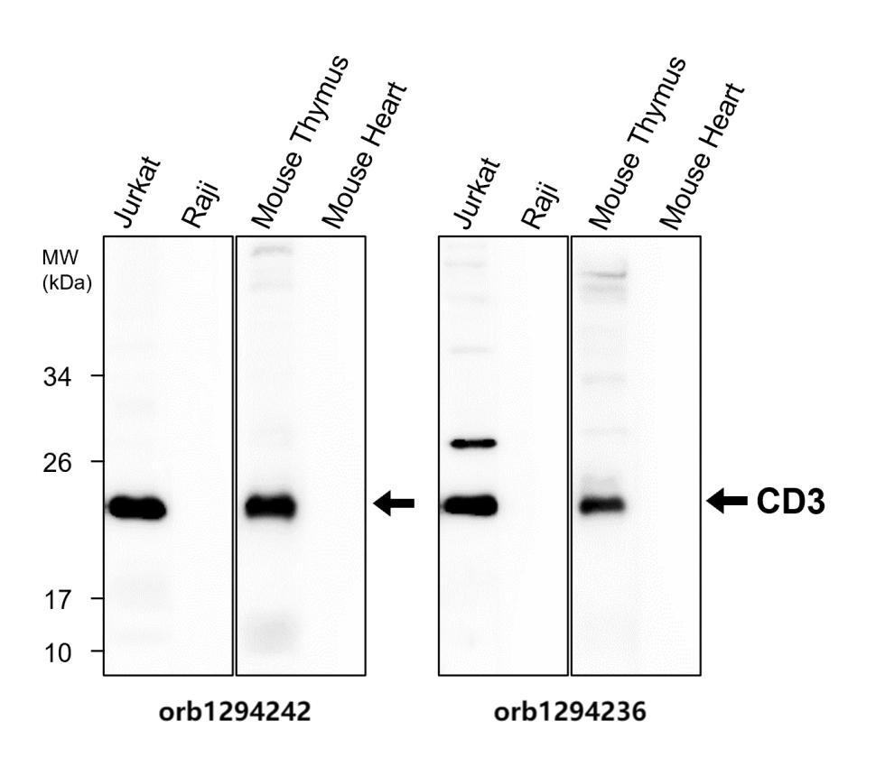
Anti-CD3 antibody at 1/1000 dilution. Lysates at 40 µg per lane. This blot was produced using a 15% SDS-PAGE. NC membrane was then blocked for an hour before being incubated with orb1294242 overnight at 4C. |
|
|
Immunofluorescence: cells were fixed with 4% paraformaldehyde for 10 min at RT, permeabilized with 0.1% NP-40 for 10 min at RT then blocked with 5% BSA for 30 min at room temperature. Cells were stained with orb1294242 anti-CD3 antibody at 1:200 and 4C. DAPI (blue) was used as the nuclear counter stain. |
|
|
Immunohistochemical analysis of paraffin embedded Human Hepatocellular cancer tissue labeling CD3 with orb1294242 at 1/100. |
|
|
Immunohistochemical analysis of paraffin embedded Human Tonsil carcinoma tissue labeling CD3 with orb1294242 at 1/200. |
|
|
Immunohistochemical analysis of paraffin embedded mouse spleen labeling CD3 with orb1294242 at 1/200 RT 1hr. Epitope Retrieval methods: Citrate Buffer, pH 6.0. |
|
|
Immunohistochemical analysis of paraffin embedded mouse spleen labeling CD3 with orb1294242 at 1/200 RT 1hr. Epitope Retrieval methods: Citrate Buffer, pH 6.0. |
|
|
Immunohistochemical analysis of paraffin embedded mouse spleen labeling CD3 with orb1294242 at 1/200 RT 1hr. Epitope Retrieval methods: Citrate Buffer, pH 6.0. |
|
|
CD3 antibody, orb1294242, Sample: Human peripheral blood, anti-CD3 antibody at 1:100. |
Product Guarantee and Expert Support
