CD antigen CD62P,CD62,CD62 antigen-like family member P,CD62P,GMP140,GMP-140,granule membrane protein 140,granule membrane protein 140kDa,granulocyte membrane protein,GRMP,LECAM3,leukocyte-endothelial cell adhesion molecule 3,PADGEM,platelet activation dependent granule-external membrane protein,platelet alpha-granule membrane protein,PSEL,P-selectin,selectin P (granule membrane protein 140kDa, antigen CD62)
Recombinant antibody was purified from cell culture supernatant
Form:
Liquid
Target:
P-Selectin/CD62P
Application Dilute:
Immunocytochemistry (ICC) 1:100 to 1:500. Epitope retrieval with citrate buffer pH 6.0 is recommended for FFPE cell sections. Immunohistochemistry (IHC) 1:100 to 1:500. Epitope retrieval with citrate buffer pH 6.0 is recommended for FFPE tissue sections.
Application Notes:
Application Notes: All western blot analysis is performed using 5% Milk-TBST for blocking and as antibody diluent. Primary antibody is incubated overnight
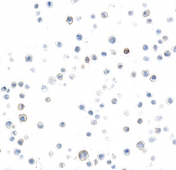
Detection of human P-Selectin/CD62P by immunocytochemistry. Sample: FFPE section of HEL 92.1.7 cells. Antibody: Rabbit anti-P-Selectin/CD62P recombinant monoclonal antibody (orb1784576). Secondary: HRP-conjugated goat anti-rabbit IgG.
Detection of human P-Selectin/CD62P by immunohistochemistry. Sample: FFPE section of tonsil. Antibody: Rabbit anti-P-Selectin/CD62P recombinant monoclonal antibody (orb1784576). Secondary: HRP-conjugated goat anti-rabbit IgG.
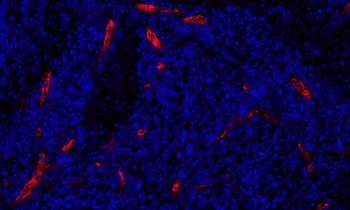
Detection of human P-Selectin/CD62P (red) by immunofluorescence. Sample: FFPE section of metastatic lymph node. Antibody: Rabbit anti-P-Selectin/CD62P recombinant monoclonal antibody (orb1784576) used at 1:100. Secondary: Invitrogen Goat anti-rabbit IgG Alexa Fluor(TM) Plus 647 1:400. Counterstain: DAPI (blue).
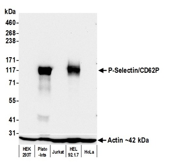
Detection of human P-Selectin/CD62P by western blot. Samples: Whole cell lysate (25 µg) from HEK293T, Platelets, Jurkat, HEL 92.1.7, and HeLa cells prepared using NETN lysis buffer. Antibody: Rabbit anti-P-Selectin/CD62P recombinant monoclonal antibody (orb1784576) used at 1:1000. Secondary: HRP-conjugated goat anti-rabbit IgG. Detection: Chemiluminescence with an exposure time of 30 seconds. Lower Panel: Rabbit anti-Actin recombinant monoclonal antibody.
* VAT and and shipping costs not included. Errors and price changes excepted