Each vial contains antibody formulated with stabilizing components, 0.9 mg NaCl, 0.2 mg Na2HPO4, and 0.05 mg NaN3. *This antibody is supplied in a stabilized formulation. Compatibility with conjugation reactions depends on the chemistry of the conjugation
Form:
Lyophilized
Target:
Parkinson disease protein 7 homolog
Application Dilute:
Western blot, 0.1-0.5µg/ml Immunohistochemistry (Paraffin-embedded Section), 0.5-1µg/ml ELISA (Cap), 1-5µg/ml
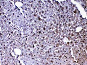
IHC analysis of PARK7 / DJ1 using anti-PARK7 / DJ1 antibody. PARK7 / DJ1 was detected in paraffin-embedded section of mouse liver tissue. Heat mediated antigen retrieval was performed in citrate buffer (pH6, epitope retrieval solution) for 20 mins. The tissue section was blocked with 10% goat serum. The tissue section was then incubated with 1 ug/ml rabbit anti-PARK7 / DJ1 Antibody overnight at 4 Biotinylated goat anti-rabbit IgG was used as secondary antibody and incubated for 30 minutes at 37 The tissue section was developed using Strepavidin-Biotin-Complex (SABC) with DAB as the chromogen.
IHC ana
IHC analysis of PARK7 / DJ1 using anti-PARK7 / DJ1 antibody. PARK7 / DJ1 was detected in paraffin-embedded section of mouse pancreas tissue. Heat mediated antigen retrieval was performed in citrate buffer (pH6, epitope retrieval solution) for 20 mins. The tissue section was blocked with 10% goat serum. The tissue section was then incubated with 1 ug/ml rabbit anti-PARK7 / DJ1 Antibody overnight at 4 Biotinylated goat anti-rabbit IgG was used as secondary antibody and incubated for 30 minutes at 37 The tissue section was developed using Strepavidin-Biotin-Complex (SABC) with DAB as the chromogen.
IHC analysis of PARK7 / DJ1 using anti-PARK7 / DJ1 antibody. PARK7 / DJ1 was detected in paraffin-embedded section of mouse testis tissue. Heat mediated antigen retrieval was performed in citrate buffer (pH6, epitope retrieval solution) for 20 mins. The tissue section was blocked with 10% goat serum. The tissue section was then incubated with 1 ug/ml rabbit anti-PARK7 / DJ1 Antibody overnight at 4 Biotinylated goat anti-rabbit IgG was used as secondary antibody and incubated for 30 minutes at 37 The tissue section was developed using Strepavidin-Biotin-Complex (SABC) with DAB as the chromogen.
IHC analysis of PARK7 / DJ1 using anti-PARK7 / DJ1 antibody. PARK7 / DJ1 was detected in paraffin-embedded section of rat liver tissue. Heat mediated antigen retrieval was performed in citrate buffer (pH6, epitope retrieval solution) for 20 mins. The tissue section was blocked with 10% goat serum. The tissue section was then incubated with 1 ug/ml rabbit anti-PARK7 / DJ1 Antibody overnight at 4 Biotinylated goat anti-rabbit IgG was used as secondary antibody and incubated for 30 minutes at 37 The tissue section was developed using Strepavidin-Biotin-Complex (SABC) with DAB as the chromogen.
IHC analysis of PARK7 / DJ1 using anti-PARK7 / DJ1 antibody. PARK7 / DJ1 was detected in paraffin-embedded section of rat pancreas tissue. Heat mediated antigen retrieval was performed in citrate buffer (pH6, epitope retrieval solution) for 20 mins. The tissue section was blocked with 10% goat serum. The tissue section was then incubated with 1 ug/ml rabbit anti-PARK7 / DJ1 Antibody overnight at 4 Biotinylated goat anti-rabbit IgG was used as secondary antibody and incubated for 30 minutes at 37 The tissue section was developed using Strepavidin-Biotin-Complex (SABC) with DAB as the chromogen.
IHC analysis of PARK7 / DJ1 using anti-PARK7 / DJ1 antibody. PARK7 / DJ1 was detected in paraffin-embedded section of rat testis tissue. Heat mediated antigen retrieval was performed in citrate buffer (pH6, epitope retrieval solution) for 20 mins. The tissue section was blocked with 10% goat serum. The tissue section was then incubated with 1 ug/ml rabbit anti-PARK7 / DJ1 Antibody overnight at 4 Biotinylated goat anti-rabbit IgG was used as secondary antibody and incubated for 30 minutes at 37 The tissue section was developed using Strepavidin-Biotin-Complex (SABC) with DAB as the chromogen.
Western blot analysis of PARK7/DJ1 using anti-PARK7/DJ1 antibody. Electrophoresis was performed on a 5-20% SDS-PAGE gel at 70V (Stacking gel) / 90V (Resolving gel) for 2-3 hours. The sample well of each lane was loaded with 30 ug of sample under reducing conditions. Lane 1: rat liver tissue lysates, Lane 2: rat RH35 whole cell lysates, Lane 3: mouse kidney tissue lysates, Lane 4: mouse liver ti
* VAT and and shipping costs not included. Errors and price changes excepted