Liquid. Purified antibody supplied in 1x PBS buffer with 0.09% (w/v) sodium azide and 2% sucrose.
Form:
Liquid. Purified antibody supplied in 1x PBS buffer with 0.09% (w/v) sodium azide and 2% sucrose.
Sequence:
Synthetic peptide located within the following region: MKIFDAAKAPIQWEERNVTAIQGPGGKWMIPSEAKESMDKNKMGLKGPLK
Target:
IDH3A
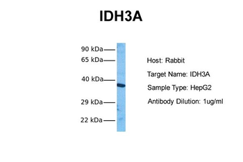
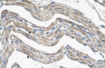
Sample Tissue: Human HepG2, Antibody Dilution: 1.0 ug/ml.
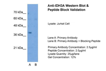
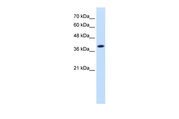
Sample Type: Jurkat, Lane A: Primary Antibody, Lane B: Primary Antibody + Blocking Peptide, Primary Antibody Concentration: 2.5 ug/ml, Peptide Concentration: 2.0 ug/ml, Lysate Quantity: 25 ug/lane, Gel Concentration: 12%.
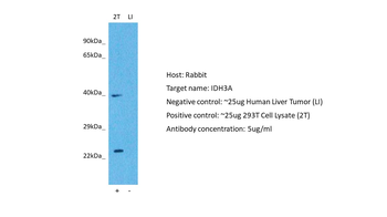
Positive control (+): 293T (2T), Negative control (-): Liver tumor (T-LI), Antibody concentration: 1 ug/ml.
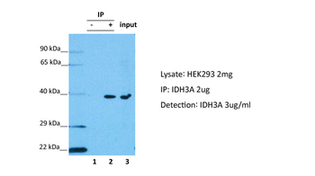
IDH3A was immunoprecipitated from 2 mg HEK293 Whole Cell Lysate with orb578394 with 1:200 dilution. Western blot was performed using orb578394 at 1/1000 dilution. Lane 1: Control IP in HEK293 Whole cell lysate. Lane 2: IDH3A IP with orb578394 in HEK293 Whole cell lysate. Lane 3: Input of HEK293 Whole cell lysate.
WB Suggested Anti-IDH3A Antibody Titration: 1.25 ug/ml, Positive Control: Jurkat cell lysate. IDH3A is strongly supported by BioGPS gene expression data to be expressed in Human Jurkat cells.
* VAT and and shipping costs not included. Errors and price changes excepted