
CANX Monoclonal Antibody, IgG2b, Clone: [15B1A7], Unconjugated, Mouse
Catalog Number:
CSB-MA004485A1M
- Images (5)

| Article Name: | CANX Monoclonal Antibody, IgG2b, Clone: [15B1A7], Unconjugated, Mouse |
| Biozol Catalog Number: | CSB-MA004485A1M |
| Supplier Catalog Number: | CSB-MA004485A1m |
| Alternative Catalog Number: | CSB-MA004485A1M-100UL, CSB-MA004485A1M-50UL |
| Manufacturer: | Cusabio |
| Host: | Mouse |
| Category: | Antikörper |
| Application: | ELISA, FC, IF, WB |
| Species Reactivity: | Human |
| Conjugation: | Unconjugated |
| Alternative Names: | Calnexin antibody, CALX_HUMAN antibody, CANX antibody, CNX antibody, FLJ26570 antibody, Histocompatibility complex class I antigen binding protein p88 antibody, IP90 antibody, Major histocompatibility complex class I antigen-binding protein p88 antibody, p90 antibody |
| Clonality: | Monoclonal |
| Clone Designation: | [15B1A7] |
| Isotype: | IgG2b |
| UniProt: | P27824 |
| Buffer: | Preservative: 0.03% Proclin 300<br />Constituents: 50% Glycerol, 0.01M PBS, PH 7.4 |
| Purity: | >95%, Protein G purified |
| Form: | Liquid |
| Target: | CANX |
| Application Dilute: | Recommended dilution: WB:1:1000-1:5000, IF:1:50-1:200, FC:1:50-1:200 |
|
|
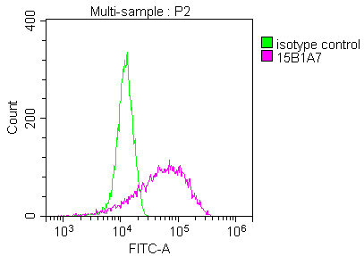
Overlay Peak curve showing Hela cells stained with CSB-MA004485A1m (red line) at 1:140. The cells were incubated in 10% normal goat serum to block non-specific protein-protein interactions followed by the antibody (1µg/1*106cells) for 1h at 4C. The secondary antibody used was FITC-conjugated Goat Anti-Mouse IgG(H+L) at 1/100 dilution for 30min at 4C. Isotype control antibody (green line) was mouse IgG1 (1µg/1*106cells) used under the same conditions. Acquisition of >10,000 events was performed. |
|
|
CSB-MA004485A1m |
|
|
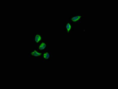
Immunofluorescence staining of Hela cells with CANX CSB-MA004485A1m at 1:140, counter-stained with DAPI. The cells were fixed in 4% formaldehyde and blocked in 10% normal Goat Serum. The cells were incubated with the antibody overnight at 4C. Nuclear DNA was labeled in blue with DAPI. The secondary antibody was FITC-conjugated AffiniPure Goat Anti-Mouse IgG (H+L). |
|
|
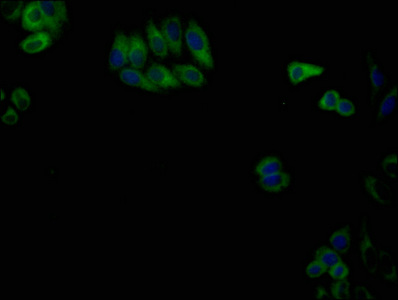
Immunofluorescence staining of HepG2 cells with CANX CSB-MA004485A1m at 1:140, counter-stained with DAPI. The cells were fixed in 4% formaldehyde and blocked in 10% normal Goat Serum. The cells were incubated with the antibody overnight at 4C. Nuclear DNA was labeled in blue with DAPI. The secondary antibody was FITC-conjugated AffiniPure Goat Anti-Mouse IgG (H+L). |
|
|
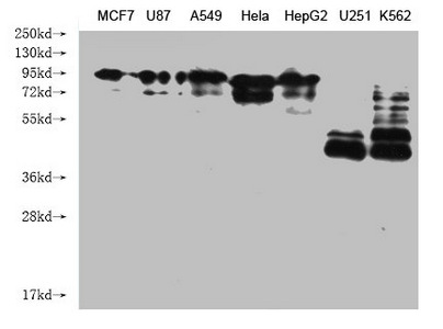
Western Blot Positive WB detected in: MCF7 whole cell lysate, U87 whole cell lysate, A549 whole cell lysate, Hela whole cell lysate, HepG2 whole cell lysate, U251 whole cell lysate, K562 whole cell lysate All lanes: CANX antibody at 1:2000 Secondary Goat polyclonal to mouse IgG at 1/50000 dilution Predicted band size: 68, 72, 56 kDa Observed band size: 90 KDa Exposure time:1min |
Product Guarantee and Expert Support
