
CIDEA Antibody, Unconjugated, Rabbit, Polyclonal
Catalog Number:
CSB-PA005431LA01HU
- Images (8)


| Article Name: | CIDEA Antibody, Unconjugated, Rabbit, Polyclonal |
| Biozol Catalog Number: | CSB-PA005431LA01HU |
| Supplier Catalog Number: | CSB-PA005431LA01HU |
| Alternative Catalog Number: | CSB-PA005431LA01HU-100UG, CSB-PA005431LA01HU-50UG |
| Manufacturer: | Cusabio |
| Host: | Rabbit |
| Category: | Antikörper |
| Application: | ELISA, IF, WB |
| Species Reactivity: | Human, Mouse |
| Conjugation: | Unconjugated |
| Alternative Names: | CIDEACell death activator CIDE-A antibody, Cell death-inducing DFFA-like effector A antibody |
| Clonality: | Polyclonal |
| UniProt: | O60543 |
| Buffer: | Liquid in PBS containing 50% glycerol, and 0.02% sodium azide. |
| Purity: | Affinity-chromatography purified |
| Form: | Liquid |
| Target: | CIDEA |
| Application Dilute: | Recommended dilution: WB:1:500-1:2000, IF:1:20-1:200 |
|
|
Im |
|
|
|
|
|
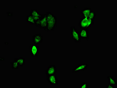
Immunofluorescent analysis of Hela cells using CSB-PA005431LA01HU at dilution of 1:100 and Alexa Fluor 488-congugated AffiniPure Goat Anti-Rabbit IgG(H+L) |
|
|
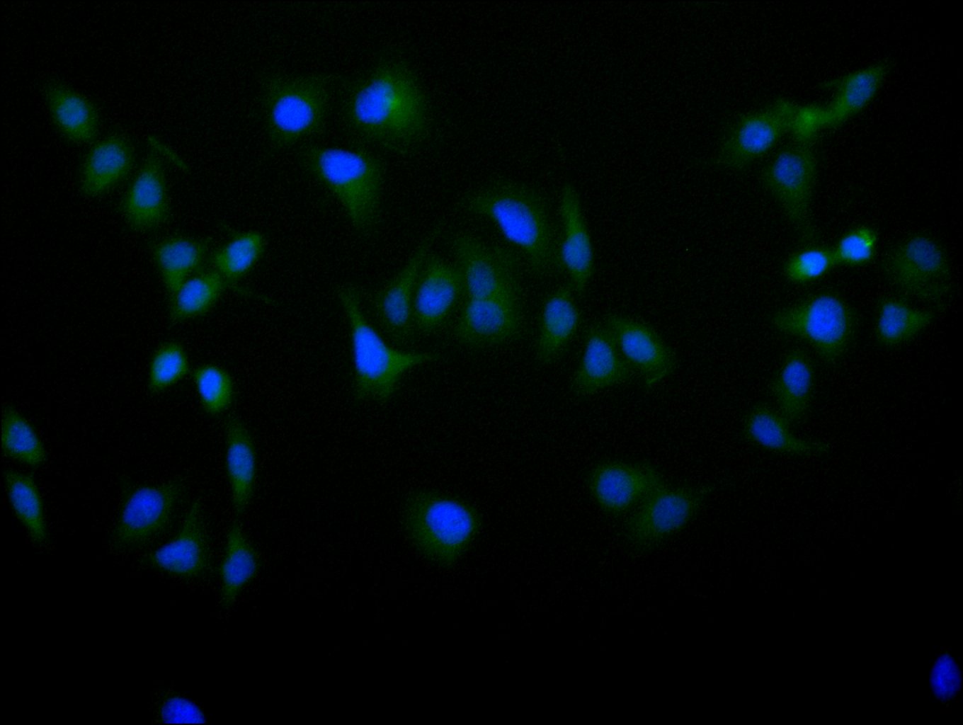
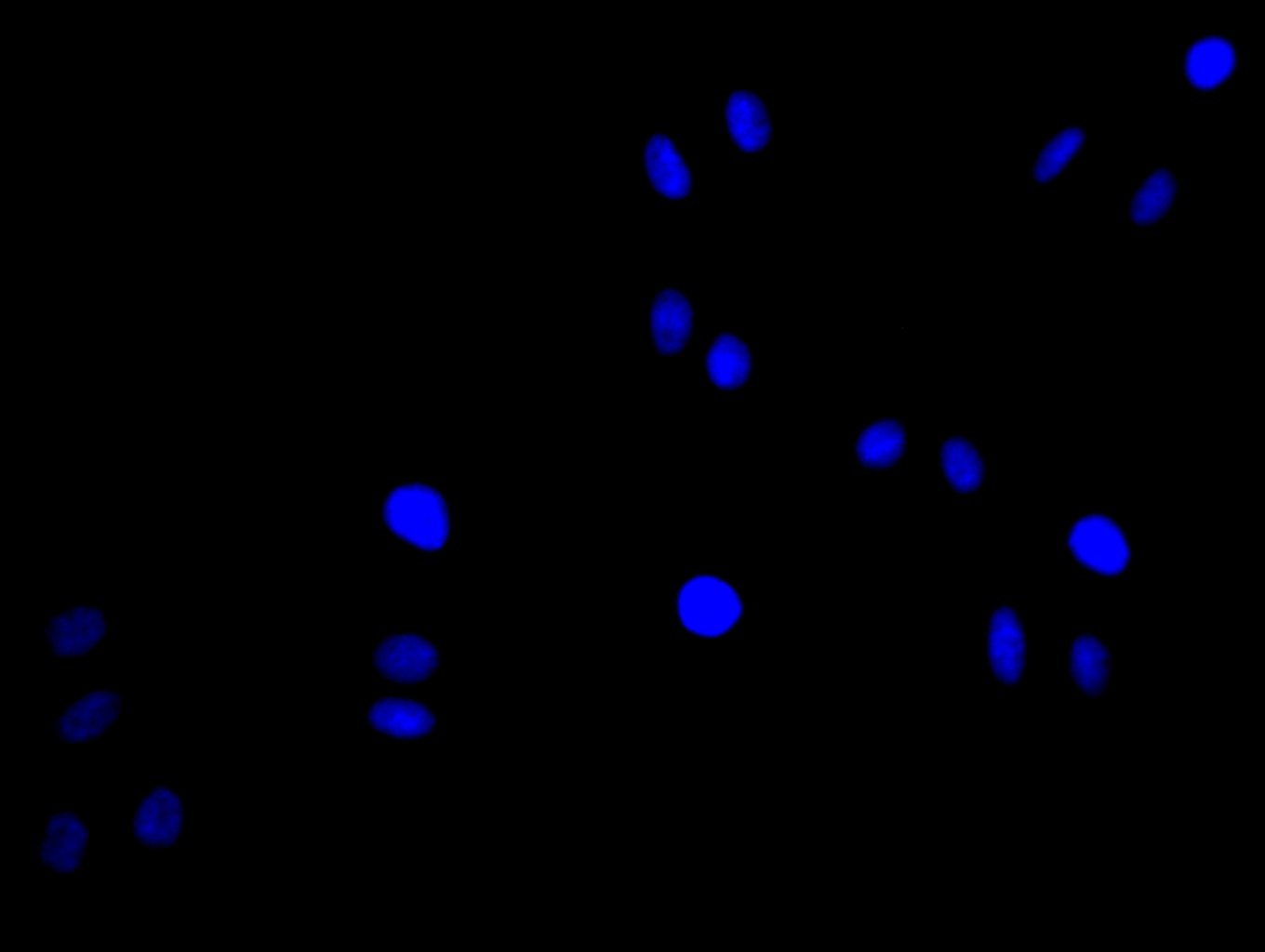
Immunofluorescence staining of Hela cell with CSB-PA005431LA01HU at 1:30, counter-stained with DAPI. The cells were fixed in 4% formaldehyde and blocked in 10% normal Goat Serum. The cells were then incubated with the antibody overnight at 4C. The secondary antibody was Alexa Fluor 488-congugated AffiniPure Goat Anti-Rabbit IgG(H+L). |
|
|
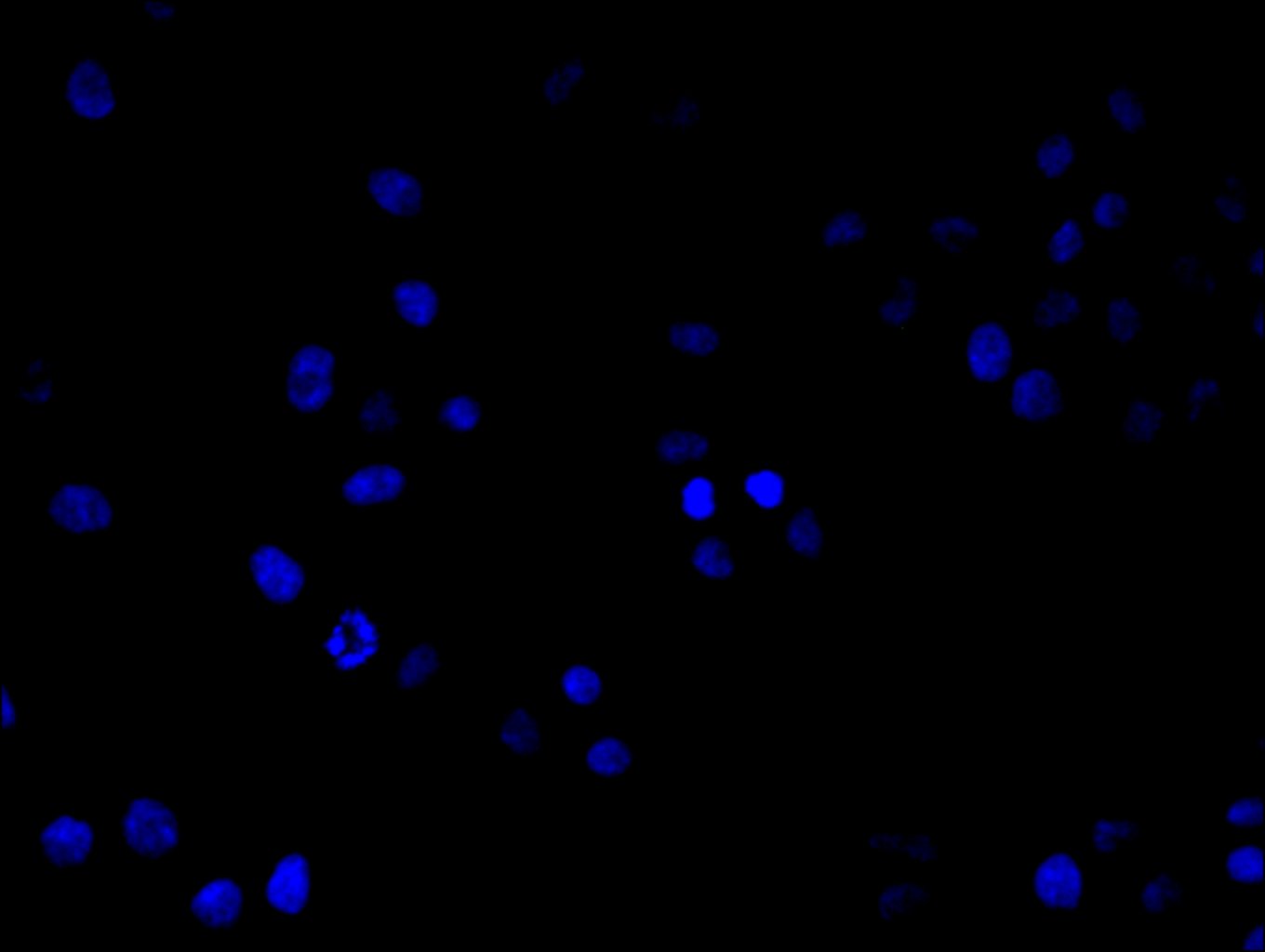
Immunofluorescence staining of Hela cell with 5% goat serum, counter-stained with DAPI. The cells were fixed in 4% formaldehyde and blocked in 10% normal Goat Serum. The cells were then incubated with the antibody overnight at 4C. The secondary antibody was Alexa Fluor 488-congugated AffiniPure Goat Anti-Rabbit IgG(H+L). |
|
|
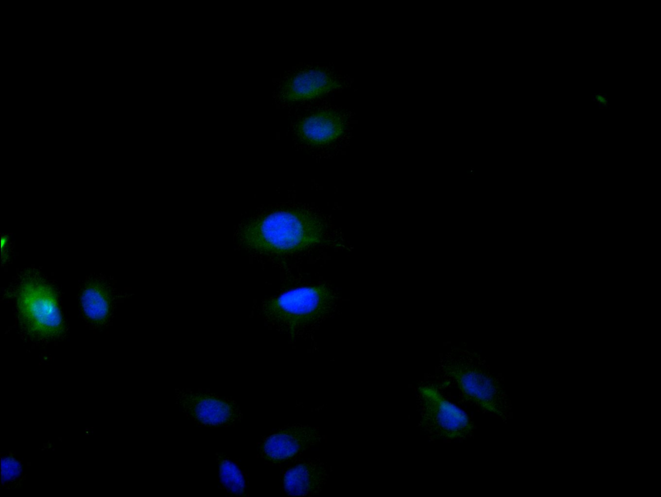
Immunofluorescence staining of A549 cell with CSB-PA005431LA01HU at 1:30, counter-stained with DAPI. The cells were fixed in 4% formaldehyde and blocked in 10% normal Goat Serum. The cells were then incubated with the antibody overnight at 4C. The secondary antibody was Alexa Fluor 488-congugated AffiniPure Goat Anti-Rabbit IgG(H+L). |
|
|
Immunofluorescence staining of A549 cell with 5% goat serum, counter-stained with DAPI. The cells were fixed in 4% formaldehyde and blocked in 10% normal Goat Serum. The cells were then incubated with the antibody overnight at 4C. The secondary antibody was Alexa Fluor 488-congugated AffiniPure Goat Anti-Rabbit IgG(H+L). |
|
|
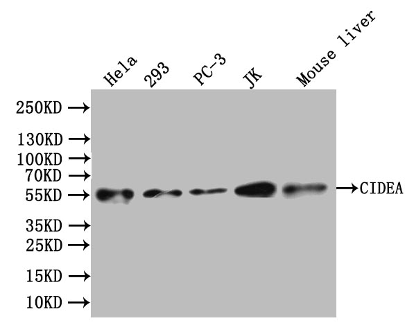
Western Blot Positive WB detected in: Hela whole cell lysate, 293 whole cell lysate, PC-3 whole cell lysate, JK whole cell lysate, Mouse Liver tissue lysate All lanes: CIDEA antibody at 1:1000 Secondary Goat polyclonal to rabbit IgG at 1/50000 dilution Predicted band size: 25 kDa Observed band size: 55 kDa |
Product Guarantee and Expert Support
