
DVL2 Antibody, Unconjugated, Rabbit, Polyclonal
Catalog Number:
CSB-PA007286ESR1HU
- Images (6)


| Article Name: | DVL2 Antibody, Unconjugated, Rabbit, Polyclonal |
| Biozol Catalog Number: | CSB-PA007286ESR1HU |
| Supplier Catalog Number: | CSB-PA007286ESR1HU |
| Alternative Catalog Number: | CSB-PA007286ESR1HU-100UL, CSB-PA007286ESR1HU-50UL |
| Manufacturer: | Cusabio |
| Host: | Rabbit |
| Category: | Antikörper |
| Application: | ELISA, IF, IHC, WB |
| Species Reactivity: | Human |
| Conjugation: | Unconjugated |
| Alternative Names: | Dishevelled 2 (homologous to Drosophila dsh) antibody, Dishevelled dsh homolog 2 antibody, dishevelled segment polarity protein 2 antibody, Dishevelled-2 antibody, Dishevelled2 antibody, DSH homolog 2 antibody, DVL 2 antibody, Dvl2 antibody, DVL2_HUMAN antibody, Segment polarity protein dishevelled homolog DVL 2 antibody, Segment polarity protein dishevelled homolog DVL-2 antibody, Segment polarity protein dishevelled homolog DVL2 antibody |
| Clonality: | Polyclonal |
| UniProt: | O14641 |
| Buffer: | PBS with 0.02% sodium azide, 50% glycerol, pH7.3. |
| Purity: | Antigen Affinity Purified |
| Form: | Liquid |
| Target: | DVL2 |
| Application Dilute: | Recommended dilution: WB:1:500-1:2000, IHC:1:20-1:500, IF:1:50-1:200 |
|
|
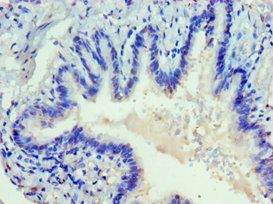
Immunohistochemistry of paraffin-embedded human lung tissue using CSB-PA007286ESR1HU at dilution of 1:100 |
|
|
Immunohistochemistry of paraffin-embedded human kidney tissue using CSB-PA007286ESR1HU at dilution of 1:100 |
|
|
Immunofluorescence staining of A549 cells with CSB-PA007286ESR1HU at 1:75, counter-stained with DAPI. The cells were fixed in 4% formaldehyde, permeabilized using 0.2% Triton X-100 and blocked in 10% normal Goat Serum. The cells were then incubated with the antibody overnight at 4°,C. The secondary antibody was Alexa Fluor 488-congugated AffiniPure Goat Anti-Rabbit IgG (H+L). |
|
|
IHC image of CSB-PA007286ESR1HU diluted at 1:227 and staining in paraffin-embedded human prostate cancer performed on a Leica BondTM system. After dewaxing and hydration, antigen retrieval was mediated by high pressure in a citrate buffer (pH 6.0). Section was blocked with 10% normal goat serum 30min at RT. Then primary antibody (1% BSA) was incubated at 4°,C overnight. The primary is detected by a biotinylated secondary antibody and visualized using an HRP conjugated SP system. |
|
|
IHC image of CSB-PA007286ESR1HU diluted at 1:227 and staining in paraffin-embedded human liver cancer performed on a Leica BondTM system. After dewaxing and hydration, antigen retrieval was mediated by high pressure in a citrate buffer (pH 6.0). Section was blocked with 10% normal goat serum 30min at RT. Then primary antibody (1% BSA) was incubated at 4°,C overnight. The primary is detected by a biotinylated secondary antibody and visualized using an HRP conjugated SP system. |
|
|
Western Blot Positive WB detected in: HepG2 whole cell lysate All lanes: DVL2 antibody at 2.3ug/ml Secondary Goat polyclonal to rabbit IgG at 1/50000 dilution Predicted band size: 79 kDa Observed band size: 79 kDa |
Product Guarantee and Expert Support
