
FBXO22 Antibody, Unconjugated, Rabbit, Polyclonal
Catalog Number:
CSB-PA854115ESR1HU
- Images (6)


| Article Name: | FBXO22 Antibody, Unconjugated, Rabbit, Polyclonal |
| Biozol Catalog Number: | CSB-PA854115ESR1HU |
| Supplier Catalog Number: | CSB-PA854115ESR1HU |
| Alternative Catalog Number: | CSB-PA854115ESR1HU-100UL, CSB-PA854115ESR1HU-50UL |
| Manufacturer: | Cusabio |
| Host: | Rabbit |
| Category: | Antikörper |
| Application: | ELISA, IHC, WB |
| Species Reactivity: | Human, Mouse |
| Conjugation: | Unconjugated |
| Alternative Names: | 0610033L19Rik antibody, 1600016C16Rik antibody, F box only protein 22 antibody, F box protein 22 antibody, F box protein FBX22p44 antibody, F-box only protein 22 antibody, F-box protein FBX22p44 antibody, FBX22 antibody, FBX22_HUMAN antibody, FBXO 22 antibody, FBXO22 antibody, FIST domain containing 1 antibody, FISTC1 antibody, FLJ13986 antibody, MGC124731 antibody, MGC31799 antibody |
| Clonality: | Polyclonal |
| UniProt: | Q8NEZ5 |
| Buffer: | Liquid in PBS containing 50% glycerol, 0.5% BSA and 0.02% sodium azide. |
| Purity: | Antigen Affinity Purified |
| Form: | Liquid |
| Target: | FBXO22 |
| Application Dilute: | Recommended dilution: WB:1:1000-1:3000, IHC:1:100-1:300 |
|
|
|
|
|
|
|
|
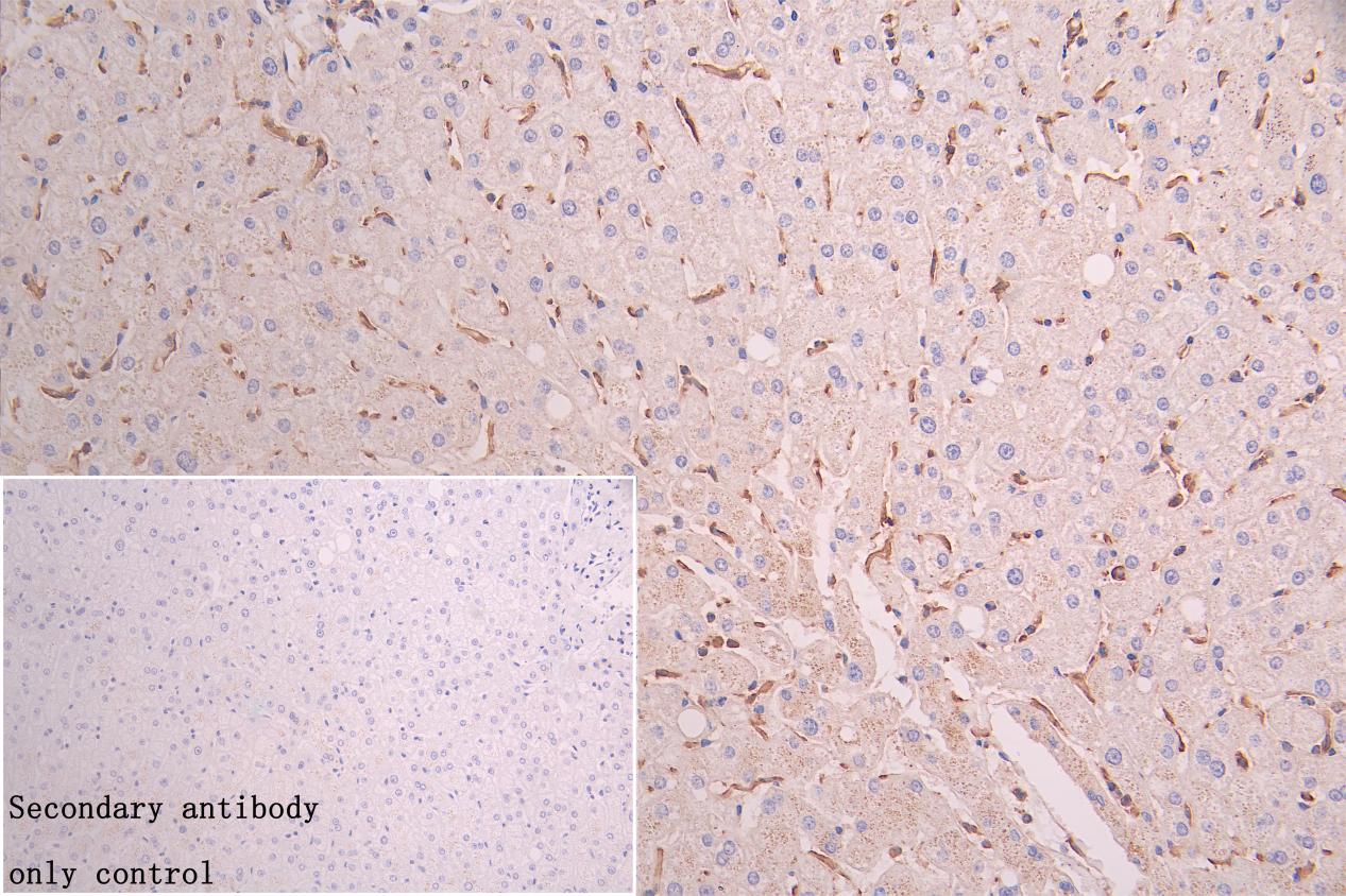
IHC image of CSB-PA854115ESR1HU diluted at 1:200 and staining in paraffin-embedded human liver tissue performed on a Leica BondTM system. After dewaxing and hydration, antigen retrieval was mediated by high pressure in a citrate buffer (pH 6.0). Section was blocked with 10% normal goat serum 30min at RT. Then primary antibody (1% BSA) was incubated at 4C overnight. The primary is detected by a Goat anti-rabbit polymer IgG labeled by HRP and visualized using 0.05% DAB.Secondary antibody only control: uses 1% BSA instead of primary antibody |
|
|
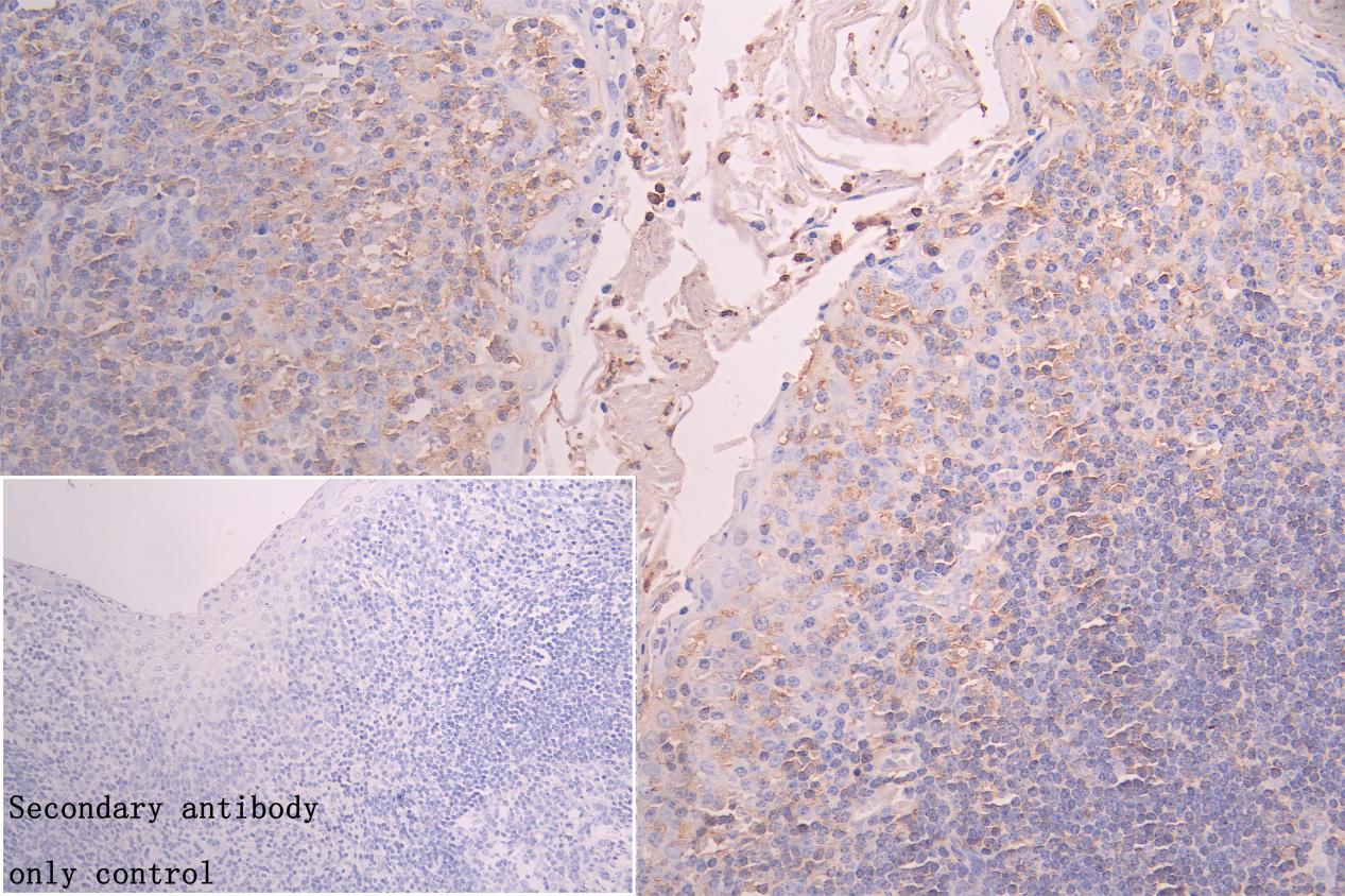
IHC image of CSB-PA854115ESR1HU diluted at 1:200 and staining in paraffin-embedded human tonsil tissue performed on a Leica BondTM system. After dewaxing and hydration, antigen retrieval was mediated by high pressure in a citrate buffer (pH 6.0). Section was blocked with 10% normal goat serum 30min at RT. Then primary antibody (1% BSA) was incubated at 4C overnight. The primary is detected by a Goat anti-rabbit polymer IgG labeled by HRP and visualized using 0.05% DAB.Secondary antibody only control: uses 1% BSA instead of primary antibody |
|
|
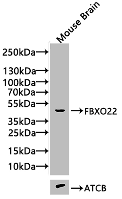
Western Blot Positive WB detected in:Mouse Brain tissue lysate(20µg) All lanes: FBXO22 antibody at 1:1000 Secondary Goat polyclonal to rabbit IgG at 1/20000 dilution Predicted band size: 45kDa Observed band size: 45 kDa Exposure time: 10s |
|
|
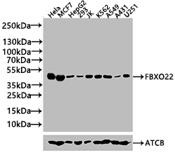
Western Blot Positive WB detected in: Hela whole cell lysate(20µg), MCF7 whole cell lysate(20µg), HepG2 whole cell lysate(20µg), 293 whole cell lysate(20µg), JK whole cell lysate(20µg), K562 whole cell lysate(20µg), A549 whole cell lysate(20µg), A431 whole cell lysate(20µg), U251 whole cell lysate(20µg) All lanes: FBXO22 antibody at 1:1000 Secondary Goat polyclonal to rabbit IgG at 1/20000 dilution Predicted band size: 45 kDa Observed band size: 45 kDa Exposure time: 10s |
Product Guarantee and Expert Support
