
AGO2 Antibody, Unconjugated, Rabbit, Polyclonal
Catalog Number:
CSB-PA891731LA01HU
- Images (5)
| Article Name: | AGO2 Antibody, Unconjugated, Rabbit, Polyclonal |
| Biozol Catalog Number: | CSB-PA891731LA01HU |
| Supplier Catalog Number: | CSB-PA891731LA01HU |
| Alternative Catalog Number: | CSB-PA891731LA01HU-100UG, CSB-PA891731LA01HU-50UG |
| Manufacturer: | Cusabio |
| Host: | Rabbit |
| Category: | Antikörper |
| Application: | ELISA, IHC, WB |
| Species Reactivity: | Human, Mouse |
| Conjugation: | Unconjugated |
| Alternative Names: | Ago 2 antibody, AGO2_HUMAN antibody, Argonaute 2 antibody, argonaute 2, RISC catalytic component antibody, Argonaute RISC catalytic component 2 antibody, Argonaute2 antibody, CTA-204B4.6 antibody, dAgo2 antibody, eIF 2C 2 antibody, eIF-2C 2 antibody, eIF2C 2 antibody, Eif2c2 antibody, Eukaryotic translation initiation factor 2C 2 antibody, Eukaryotic translation initiation factor 2C subunit 2 antibody, hAgo2 antibody, MGC3183 antibody, PAZ Piwi domain protein antibody, PPD antibody, Protein argonaute-2 antibody, Protein slicer antibody, Q10 antibody, Slicer protein antibody |
| Clonality: | Polyclonal |
| UniProt: | Q9UKV8 |
| Buffer: | Preservative: 0.03% Proclin 300<br />Constituents: 50% Glycerol, 0.01M PBS, PH 7.4 |
| Purity: | >95%, Protein G purified |
| Form: | Liquid |
| Target: | AGO2 |
| Application Dilute: | Recommended dilution: WB:1:1000-1:5000, IHC:1:100-1:1500 |
|
|
|
|
|
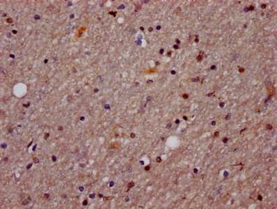
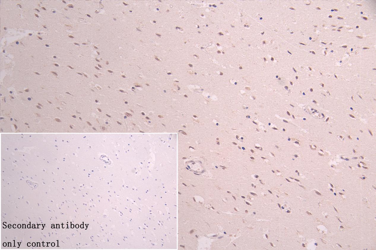
IHC image of CSB-PA891731LA01HU diluted at 1:800 and staining in paraffin-embedded human brain tissue performed on a Leica BondTM system. After dewaxing and hydration, antigen retrieval was mediated by high pressure in a citrate buffer (pH 6.0). Section was blocked with 10% normal goat serum 30min at RT. Then primary antibody (1% BSA) was incubated at 4C overnight. The primary is detected by a Goat anti-rabbit polymer IgG labeled by HRP and visualized using 0.05% DAB.Secondary antibody only control: uses 1% BSA instead of primary antibody |
|
|
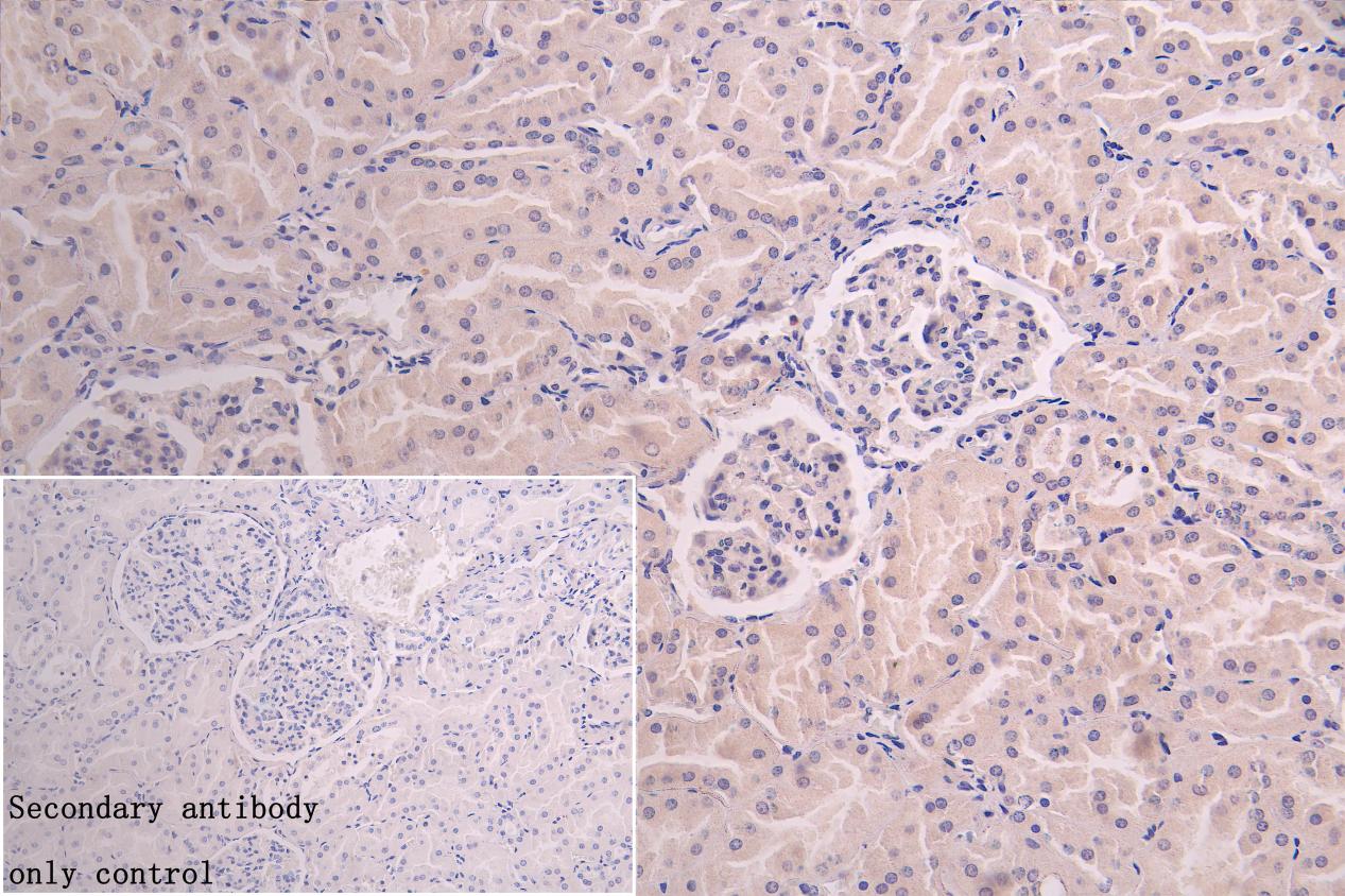
IHC image of CSB-PA891731LA01HU diluted at 1:800 and staining in paraffin-embedded human kidney tissue performed on a Leica BondTM system. After dewaxing and hydration, antigen retrieval was mediated by high pressure in a citrate buffer (pH 6.0). Section was blocked with 10% normal goat serum 30min at RT. Then primary antibody (1% BSA) was incubated at 4C overnight. The primary is detected by a Goat anti-rabbit polymer IgG labeled by HRP and visualized using 0.05% DAB.Secondary antibody only control: uses 1% BSA instead of primary antibody |
|
|
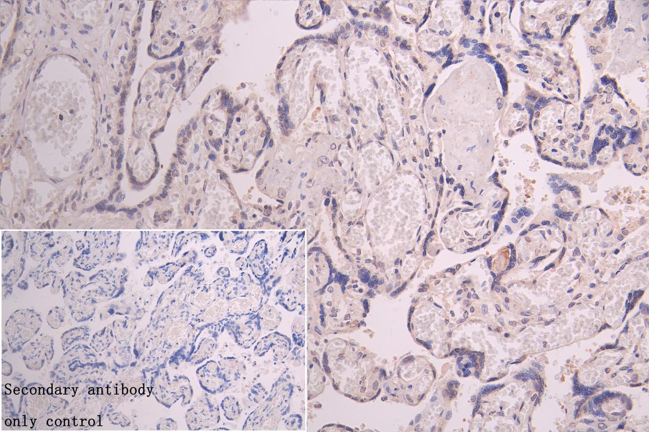
IHC image of CSB-PA891731LA01HU diluted at 1:800 and staining in paraffin-embedded human placenta tissue performed on a Leica BondTM system. After dewaxing and hydration, antigen retrieval was mediated by high pressure in a citrate buffer (pH 6.0). Section was blocked with 10% normal goat serum 30min at RT. Then primary antibody (1% BSA) was incubated at 4C overnight. The primary is detected by a Goat anti-rabbit polymer IgG labeled by HRP and visualized using 0.05% DAB.Secondary antibody only control: uses 1% BSA instead of primary antibody |
|
|
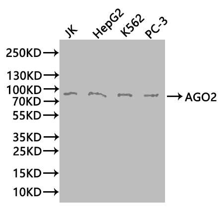
Western Blot Positive WB detected in: JK whole cell lysate(20µg), HepG2 whole cell lysate(20µg), K562 whole cell lysate(20µg), PC-3 whole cell lysate(20µg) All lanes: AGO2 antibody at 1:1000 Secondary Goat polyclonal to rabbit IgG at 1/50000 dilution Predicted band size: 98 kDa Observed band size: 98 kDa Exposure time:1min |
Product Guarantee and Expert Support
