
eIF3e antibody, Unconjugated, Rabbit, Polyclonal
Catalog Number:
GTX112342
- Images (5)
| Article Name: | eIF3e antibody, Unconjugated, Rabbit, Polyclonal |
| Biozol Catalog Number: | GTX112342 |
| Supplier Catalog Number: | GTX112342 |
| Alternative Catalog Number: | GTX112342-100,GTX112342-25 |
| Manufacturer: | GeneTex |
| Host: | Rabbit |
| Category: | Antikörper |
| Application: | ICC, IHC-P, WB |
| Species Reactivity: | Human, Mouse |
| Immunogen: | Recombinant protein encompassing a sequence within the center region of human eIF3e. The exact sequence is proprietary. |
| Conjugation: | Unconjugated |
| Alternative Names: | eukaryotic translation initiation factor 3 subunit E , EIF3-P48 , EIF3S6 , INT6 , eIF3-p46 |
| Application Notes: | WB: 1:500-1:3000. ICC/IF: 1:100-1:1000. IHC-P: 1:100-1:1000. *Optimal dilutions/concentrations should be determined by the researcher.Not tested in other applications. |
|
|
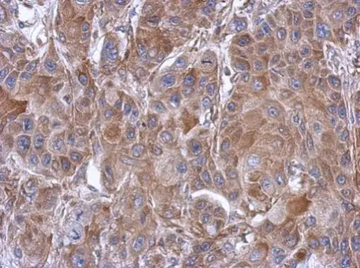
GTX112342 IHC-P Image |
|
|
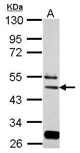
Sample (30 ug of whole cell lysate) A: BCL-1 10% SDS PAGE GTX112342 diluted at 1:2000 |
|
|
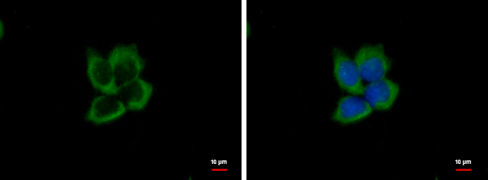
eIF3e antibody detects EIF3E protein at cytoplasm by immunofluorescent analysis. Sample: A431 cells were fixed in ice-cold MeOH for 5 min. Green: EIF3E protein stained by eIF3e antibody (GTX112342) diluted at 1:500. Blue: Hoechst 33342 staining. |
|
|
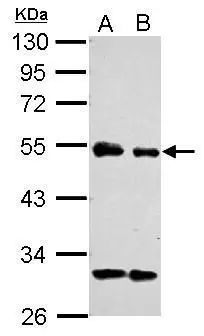
Sample (30 ug of whole cell lysate) A: Jurkat B: Raji 10% SDS PAGE GTX112342 diluted at 1:2000 |
|
|
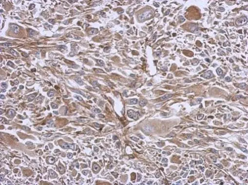
Immunohistochemical analysis of paraffin-embedded C2C12 xenograft, using eIF3e(GTX112342) antibody at 1:500 dilution. Antigen Retrieval: Trilogy™ (EDTA based, pH 8.0) buffer, 15min |
Product Guarantee and Expert Support
