
ATP citrate lyase antibody [N1N2], N-term, Unconjugated, Rabbit, Polyclonal
Catalog Number:
GTX112387
- Images (5)
| Article Name: | ATP citrate lyase antibody [N1N2], N-term, Unconjugated, Rabbit, Polyclonal |
| Biozol Catalog Number: | GTX112387 |
| Supplier Catalog Number: | GTX112387 |
| Alternative Catalog Number: | GTX112387-100,GTX112387-25 |
| Manufacturer: | GeneTex |
| Host: | Rabbit |
| Category: | Antikörper |
| Application: | ICC, IHC-P, WB |
| Species Reactivity: | Human, Mouse, Rat |
| Immunogen: | Recombinant protein encompassing a sequence within the N-terminus region of human ATP citrate lyase. The exact sequence is proprietary. |
| Conjugation: | Unconjugated |
| Alternative Names: | ATP citrate lyase , ACL , ATPCL , CLATP |
| Application Notes: | WB: 1:1000-1:10000. ICC/IF: 1:100-1:1000. IHC-P: 1:100-1:1000. *Optimal dilutions/concentrations should be determined by the researcher.Not tested in other applications. |
|
|
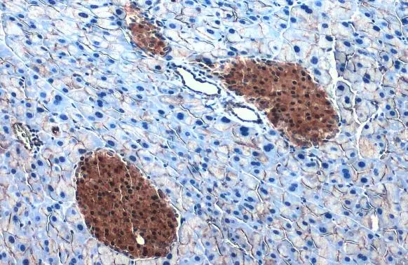
GTX112387 IHC-P Image |
|
|
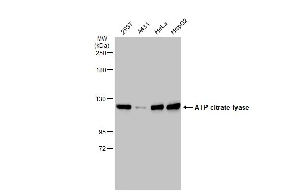
Various whole cell extracts (30 μg) were separated by 5% SDS-PAGE, and the membrane was blotted with ATP citrate lyase antibody [N1N2], N-term (GTX112387) diluted at 1:5000. The HRP-conjugated anti-rabbit IgG antibody (GTX213110-01) was used to detect the primary antibody. |
|
|
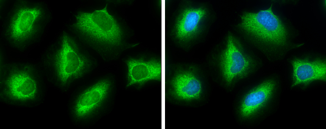
ATP citrate lyase antibody [N1N2], N-term detects ATP citrate lyase protein at cytoplasm by immunofluorescent analysis. Sample: HeLa cells were fixed in ice-cold MeOH for 5 min. Green: ATP citrate lyase protein stained by ATP citrate lyase antibody [N1N2], N-term (GTX112387) diluted at 1:500. Blue: Hoechst 33342 staining. |
|
|
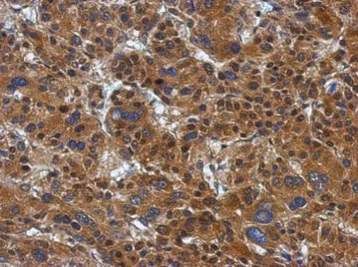
Immunohistochemical analysis of paraffin-embedded Hepatoma, using ATP citrate lyase(GTX112387) antibody at 1:500 dilution. Antigen Retrieval: Trilogy™ (EDTA based, pH 8.0) buffer, 15min |
|
|
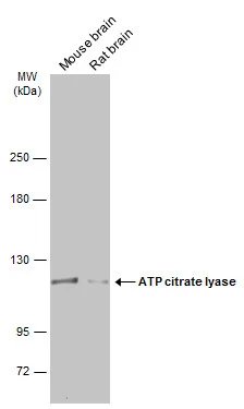
Various tissue extracts (50 μg) were separated by 5% SDS-PAGE, and the membrane was blotted with ATP citrate lyase antibody [N1N2], N-term (GTX112387) diluted at 1:5000. The HRP-conjugated anti-rabbit IgG antibody (GTX213110-01) was used to detect the primary antibody. |
Product Guarantee and Expert Support
