
LTA4H antibody [9C5], IgG2b, Unconjugated, Mouse, Monoclonal
Catalog Number:
GTX84176
- Images (9)

| Article Name: | LTA4H antibody [9C5], IgG2b, Unconjugated, Mouse, Monoclonal |
| Biozol Catalog Number: | GTX84176 |
| Supplier Catalog Number: | GTX84176 |
| Alternative Catalog Number: | GTX84176-100 |
| Manufacturer: | GeneTex |
| Host: | Mouse |
| Category: | Antikörper |
| Application: | IHC-P, IP, WB |
| Species Reactivity: | Canine, Human, Monkey, Mouse |
| Immunogen: | Full-length protein expressed in 293T cell transfected with human LTA4H expression vector |
| Conjugation: | Unconjugated |
| Alternative Names: | leukotriene A4 hydrolase |
| Hydrolyzes an epoxide moiety of leukotriene A4 (LTA-4) to form leukotriene B4 (LTB-4). The enzyme also has some peptidase activity. |
| Clonality: | Monoclonal |
| Concentration: | 1 mg/ml (Please refer to the vial label for the specific concentration.) |
| Clone Designation: | [9C5] |
| Molecular Weight: | 69 |
| Isotype: | IgG2b |
| NCBI: | 4048 |
| UniProt: | P09960 |
| Buffer: | PBS pH7.3, 1% BSA, 50% Glycerol, 0.02% Sodium azide. |
| Source: | Human |
| Purity: | Purified by affinity chromatography |
| Form: | Liquid |
| Application Notes: | WB: 1:200. IHC-P: 1:100-1:200. *Optimal dilutions/concentrations should be determined by the researcher.Not tested in other applications. |
|
|
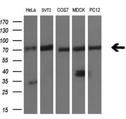
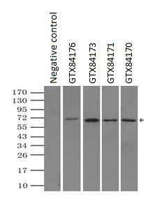
WB analysis of various cell lines using GTX84176 LTA4H antibody [9C5]. Loading : 10 ug per lane Dilution : 1:200 |
|
|
GTX84176 WB Image |
|
|
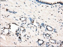
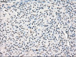
IHC-P analysis of pancreas tissue using GTX84176 LTA4H antibody [9C5]. Antigen retrieval : Heat-induced epitope retrieval by 10mM citrate buffer, pH6.0, 100ºC for 10min. Dilution : 1:50 |
|
|
IHC-P analysis of endometrium tissue using GTX84176 LTA4H antibody [9C5]. Antigen retrieval : Heat-induced epitope retrieval by 10mM citrate buffer, pH6.0, 100ºC for 10min. Dilution : 1:50 |
|
|
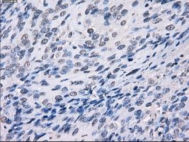
IHC-P analysis of bladder carcinoma tissue using GTX84176 LTA4H antibody [9C5]. Antigen retrieval : Heat-induced epitope retrieval by 10mM citrate buffer, pH6.0, 100ºC for 10min. Dilution : 1:50 |
|
|
IHC-P analysis of colon tissue using GTX84176 LTA4H antibody [9C5]. Antigen retrieval : Heat-induced epitope retrieval by 10mM citrate buffer, pH6.0, 100ºC for 10min. Dilution : 1:50 |
|
|
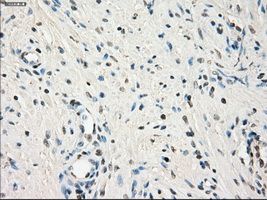
IHC-P analysis of prostate tissue using GTX84176 LTA4H antibody [9C5]. Antigen retrieval : Heat-induced epitope retrieval by 10mM citrate buffer, pH6.0, 100ºC for 10min. Dilution : 1:50 |
|
|
IP analysis of DDDDK tagged LTA4H overexpressed HEK293T lysate using GTX84176 LTA4H antibody [9C5].After extensive wash to remove any non-specific binding, the immuno-precipitated products were analyzed with rabbit anti-DDDDK polyclonal antibody. IP reaction : 2μg antibody / 500μl cell lysate |
|
|
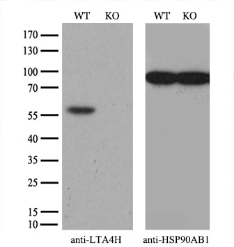
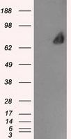
WB analysis of HEK293T cells transfected with LTA4H plasmid (Right) or empty vector (Left) for 48 hrs using GTX84176 LTA4H antibody [9C5]. Loading : 5 ug per lane |
Product Guarantee and Expert Support
