
beta Catenin Antibody, KO Validated, Unconjugated, Rabbit, Polyclonal
Catalog Number:
PRS-13-991
- Images (9)

| Article Name: | beta Catenin Antibody, KO Validated, Unconjugated, Rabbit, Polyclonal |
| Biozol Catalog Number: | PRS-13-991 |
| Supplier Catalog Number: | 13-991 |
| Alternative Catalog Number: | PRS-13-991-100 |
| Manufacturer: | ProSci |
| Host: | Rabbit |
| Category: | Antikörper |
| Application: | IHC, IP, WB |
| Species Reactivity: | Human, Mouse, Rat |
| Immunogen: | A synthetic peptide corresponding to a sequence within amino acids 650-750 of human beta Catenin (NP_001895.1). |
| Conjugation: | Unconjugated |
| Alternative Names: | CTNNB1, CTNNB, MRD19, armadillo |
| Application Dilute: | Optimal dilutions for each application to be determined by the researcher. |
| Application Notes: | WB: 1:500 - 1:2000IHC: 1:50 - 1:200IP: 1:50 - 1:200 |
|
|
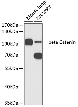
Western blot analysis of extracts of various cell lines, using beta Catenin antibody (13-991) at 1:1000 dilution. Secondary antibody: HRP Goat Anti-Rabbit IgG (H+L) at 1:10000 dilution. Lysates/proteins: 25ug per lane. Blocking buffer: 3% nonfat dry milk in TBST. Detection: ECL Basic Kit. Exposure time: 90s. |
|
|
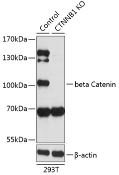
Western blot analysis of extracts from normal (control) and beta Catenin knockout (KO) 293T cells, using beta Catenin antibody (13-991) at 1:1000 dilution. Secondary antibody: HRP Goat Anti-Rabbit IgG (H+L) at 1:10000 dilution. Lysates/proteins: 25ug per lane. Blocking buffer: 3% nonfat dry milk in TBST. Detection: ECL Basic Kit. Exposure time: 90s. |
|
|
Immunohistochemistry of paraffin-embedded rat brain using beta Catenin antibody (13-991) at dilution of 1:100 (40x lens). |
|
|
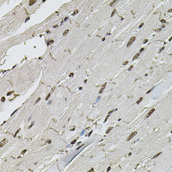
Immunohistochemistry of paraffin-embedded rat heart using beta Catenin antibody (13-991) at dilution of 1:100 (40x lens). |
|
|
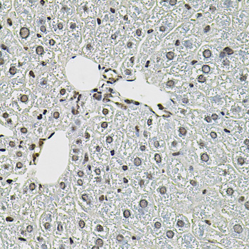
Immunohistochemistry of paraffin-embedded mouse liver using beta Catenin antibody (13-991) at dilution of 1:100 (40x lens). |
|
|
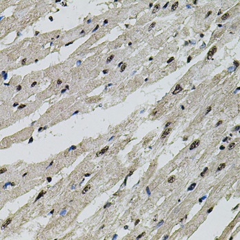
Immunohistochemistry of paraffin-embedded mouse heart using beta Catenin antibody (13-991) at dilution of 1:100 (40x lens). |
|
|
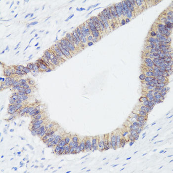
Immunohistochemistry of paraffin-embedded rat fallopian tube using beta Catenin antibody (13-991) at dilution of 1:150 (40x lens). |
|
|
Immunohistochemistry of paraffin-embedded human colon carcinoma using beta Catenin antibody (13-991) at dilution of 1:150 (40x lens). |
|
|
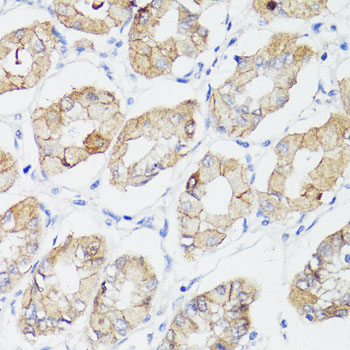
Immunohistochemistry of paraffin-embedded human stomach using beta Catenin antibody (13-991) at dilution of 1:150 (40x lens). |
Product Guarantee and Expert Support
