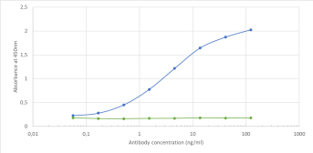
Binding curve of anti-CD155 antibody 3F1 (24-687) to recombinant mouse CD155 Fc-Fusion Protein. ELISA Plate coated with recombinant mouse CD155 Fc-Fusion Protein (Pr00231-1.9, Antibody) at a concentration of 5 ug/ml. A 3-fold serial dilution from 10-000 ng/ml was performed using 24-687. For detection- a 1:4000 dilution of HRP-lnti-ribody was used.
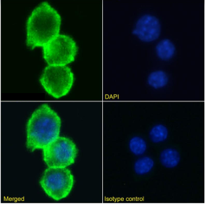
Immunofluorescence staining of RAW 264.7 cells using anti-CD155 antibody (3F1. Immunofluorescence analysis of paraformaldehyde fixed RAW 264.7 cells stained with the chimeric r version of 3F1 (24-687) at 10 ug/ml followed by Alexa Fluor 488 secondary antibody (2 ug/ml)- showing membrane staining. The nuclear stain is DAPI (blue). Panels show from left-right- top-bottom 24-687- DAPI- merged channels and an isotype control. The isotype control was stained with anti-Fluorescein antibody (3.0) followed by Alexa Fluor 488 secondary antibody.
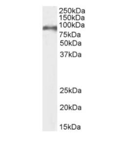
Western Blot using anti-CD155 antibody 3F1 ( Mouse thymus lysate (35ug protein in RIPA buffer) was resolved on an SDS PAGE gel and blots probed with the chimeric r version of 3F1 (24-687) at 0.1 ug/ml before detection using an anti-rondary antibody. A primary incubation of 1h was used and protein was detected by chemiluminescence.
* VAT and and shipping costs not included. Errors and price changes excepted