
Mouse IgG (H&L) Antibody DyLight(TM) 680 Conjugated Pre-Adsorbed, DL680, Donkey, Polyclonal
Catalog Number:
ROC-610-744-124
- Images (8)
| Article Name: | Mouse IgG (H&L) Antibody DyLight(TM) 680 Conjugated Pre-Adsorbed, DL680, Donkey, Polyclonal |
| Biozol Catalog Number: | ROC-610-744-124 |
| Supplier Catalog Number: | 610-744-124 |
| Alternative Catalog Number: | ROC-610-744-124 |
| Manufacturer: | Rockland Immunochemicals |
| Host: | Donkey |
| Category: | Antikörper |
| Application: | DOT, ELISA |
| Species Reactivity: | Mouse |
| Immunogen: | Mouse IgG whole molecule |
| Conjugation: | DL680 |
| Alternative Names: | Donkey anti-Mouse IgG DyLight 680(TM) Conjugated Antibody, Donkey anti Mouse IgG Antibody DyLight 680(TM) Conjugation |
| Clonality: | Polyclonal |
| Concentration: | 1.0 mg/mL by UV absorbance at 280 nm |
| Buffer: | 0.02 M Potassium Phosphate, 0.15 M Sodium Chloride, pH 7.2 |
| Form: | Lyophilized |
| Target: | Mouse |
| Antibody Type: | Secondary Antibody |
| Application Dilute: | FLISA: >1:20,000, IF Microscopy: >1:5,000, WB: >1:10,000 |
| Application Notes: | Anti-Mouse IgG DyLight680 Antibody has been tested by ELISA and dot blot and is designed for immunofluorescence microscopy, fluorescence based plate assays (FLISA) and fluorescent western blotting. This product is also suitable for multiplex analysis, in |
|
|
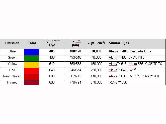
Properties of DyLight(TM) Conjugates. |
|
|
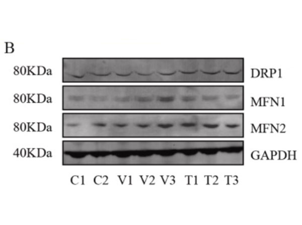
Assessment of mitochondrial fusion and fission. B. Representative western blots (original blots are shown in supplementary Fig. S10) and quantification of MFN1/2 and DRP1. No significant changes in the relative levels of proteins that facilitate mitochondrial fusion (MFN1/2) and fission (DRP1) between non-disease (control) and mutant primary fibroblasts. Data are depicted as mean SD, n = 3. The primary antibodies used as follows: MFN1 1:400, MFN2 ( 1:400, DRP1 1:100 and GAPDH 1:30,000 dilutions overnight at 4 C. The membranes were then incubated with fluorescent conjugated secondary antibodies for 1 h, DyLight 800 conjugated goat Anti-Rabbit IgG (611-145-002), Antibody DyLight 680 conjugated Anti-Rabbit IgG made in goat (611-144-003), DyLight 800 conjugated goat Anti-Mouse IgG (610-145-002), and DyLight 680 conjugated donkey Anti-Mouse IgG (610-744-124). Fig 3. PMID: 33725513. |
|
|
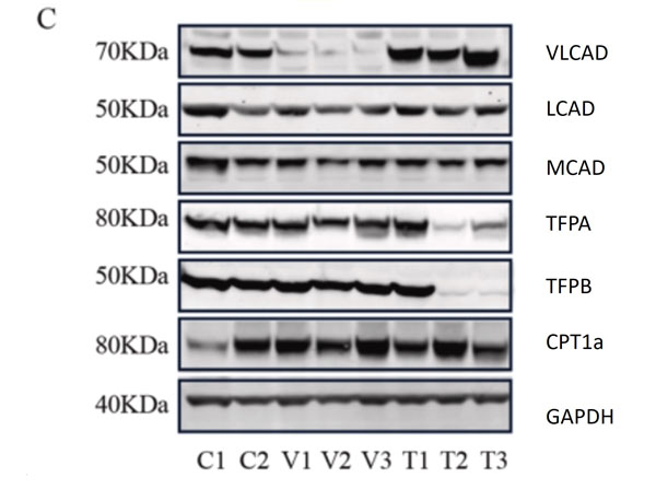
C. Representative western blots, original blots are shown in (supplementary Fig S8-9). And densitometric quantification of relative protein levels from western blots. Data are depicted as mean SD, n = 3, **P < 0.01, ***P < 0.001 and ****P < 0.0001 by one-way ANOVA. Intracellular transport, activation, mitochondrial transport, beta-oxidation, carnitine shuttle, and auxiliary proteins. The primary antibodies used as follows: VLCAD 1:1000, MCAD 1:1000, LCAD 1:1000, TFPa 1:500, TFPb 1:3000, CPT1alpha 1:1000, and GAPDH 1:30,000 dilutions overnight at 4 C. The membranes were then incubated with fluorescent conjugated secondary antibodies for 1 h, DyLight 800 conjugated goat Anti-Rabbit IgG (611-145-002), DyLight 680 conjugated goat Anti-Rabbit IgG (611-144-003), DyLight 800 conjugated goat Anti-Mouse IgG (610-145-002), and DyLight 680 conjugated donkey Anti-Mouse IgG (610-744-124). Fig 1. PMID: 33725513. |
|
|
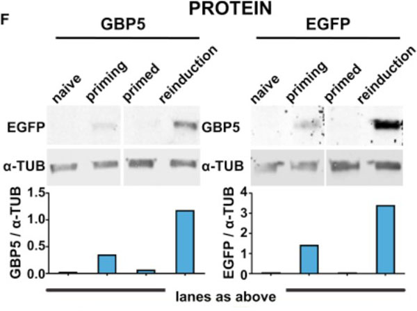
Priming Results in Increased Frequency of Activation and EnhancedGBP5Expression upon Reinduction.(F) EGFP::GBP5 cells were subjected to the IFNgamma treatment regimen outlined inFigure1B, processed for fluorescence western blotting, and probed for GBP5 and EGFP expression. alpha-TUB, loading control. Tubulin-normalized fluorescence intensities are plotted. Fig 3. PMID: 33108759. |
|
|
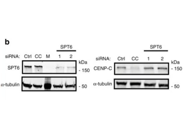
Depletion of human SPT6 leads to the loss of CENP-A maintenance. HeLa cells expressing SNAP-tagged CENP-A were treated with TMR-star to detect previously incorporated CENP-A and siRNA-treated to deplete proteins indicated in (b,c). Cells were then synchronized in S phase by a thymidine block and released. Cells were allowed transit through G1 phase and were collected at the next G1/S boundary by re-addition of thymine.bCells were treated with indicated siRNAs for 48h and extracts were processed for immunoblotting and probed with indicated antibodies. CC CENP-C, M Marker.N=3 independent experiments. Fig 6. PMID: 32522980. |
|
|
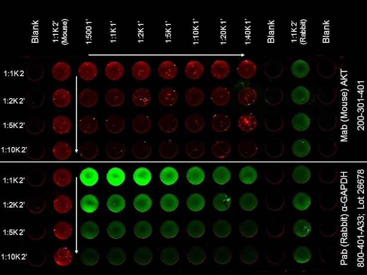
ELISA of DyLight(TM) 680 Conjugated Donkey Anti-Mouse Secondary Antibody. Antigen: HCT-116 cell line. Coating amount: Confluent in the 96 well plate. Primary antibody: AKT or GAPDH antibody at 2 µg/mL. Dilution series: Primary and Secondary Antibodies 2-fold. Mid-point concentration: N/A. Secondary antibody: DyLight(TM) 680 donkey secondary antibody and DyLight(TM) 800 goat secondary antibody starting at 1:1,000. Substrate: None. |
|
|
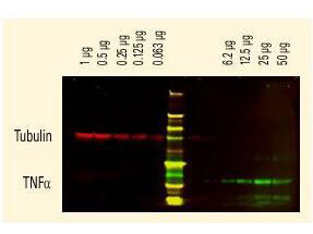
DyLight(TM) dyes can be used for two-color Western Blot detection with low background and high signal. Anti-tubulin was detected using a DyLight(TM) 680 conjugate. Anti-TNFa was detected using a DyLight(TM) 800 conjugate. The image was captured using the Odyssey Infrared Imaging System developed by LI-COR. |
|
|
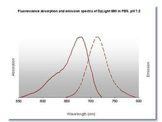
DyLight(TM) 680 Fluorescence Spectra. |
Product Guarantee and Expert Support
