COVID-19 immunity
Cytometric lymphocyte detection
Basis of successfull vaccinations against pathogens as the Coronavirus SARS-CoV-2 is the type, and efficiency of the induced immune response. The human adaptive immune system relies on both, the humoral immunity as well as the T-cell mediated immunity. Our tools for flow cytometrical analysis of B- and T-cell subsets allow to study the underlying processes of the vaccine or infection induced immunity.
In case of SARS-CoV-2, the longterm immunity is heterogeneous in patients, and involves, beside antibodies, memory B-cells, both CD8+ T cells, and CD4+ T cells (Cox et al.. 2020; Dan et al., 2021). Coronavirus vaccine development has so far largely focused on antibodies, as those are promising to provide ‘sterilizing immunity’, which not only reduces the severity of an illness, but prevents infection. However, fastspreading new COVID-19 variants, suspected to to be resistant to antibodies, put T-cell mediated immunity anew in the spotlight (Nature News, 2021). Also do studies on other coronaviruses as SARS-CoV-1 and MERS support the hope of a stable longterm T-cell immunity (Sariol et al., 2020). The leading three COVID-19 vaccines induce both types of immunity (BMJ 2020), and a novel vaccine is in development at the Tübingen University, designed to activate specifically T-cell responses.
Still, there remain open questions regarding the T-cell immunity to SARS-CoV-2, like which viral factors regulate the strength and efficacy of the T-cell response, and do these factors influence the severity of COVID-19, or what type of T-cell response elicited by vaccines will be the best predictor of protection from disease? (Karlsson et al., 2020)
We support the important research on T-cell immunity in COVID-19 with a range of products for cytometrical analysis of B- and T-cell subsets.
COVID-19 EXBIO Dry Reagent kits
With the Exbio Dry Reagent Kits, we can provide you with an efficient, reliable and fast tool for analyzing carefully selected cellular subpopulations that are important for characterizing the patient's immune system status, monitoring it over time, monitoring the impact of medical intervention and providing information helpul in determination of patient‘s prognosis.

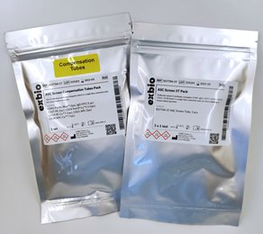
B-lymphocyte subsets: DryFlowEx ASC Screen Kit (EXB-ED7704):
Identification and percentage enumeration of differentiated B-lymphocyte subsets secreting antibodies (ASC – Antibody Secreting Cells) after virus infection in EDTA anticoagulated peripheral blood using Flow Cytometry. The ED7704 DryFlowEx ASC Screen Kit is a multicolor panel of antibody conjugates CD45 Pacific Blue™ / IgD FITC / CD27 PE / CD24 PerCP-Cy™5.5 / CD19 PE-Cy™7 / CD21 APC / CD38 APC-Cy™7 dried in a single flow cytometry tube for direct labelling of human blood specimen.
T-lymphocyte subsets: DryFlowEx ACT T Screen Kit (EXB-ED7705):
Identification and percentage enumeration of activated T-lymphocyte subsets and a specific subset of Follicular Helper T lymphocytes (Tfh) after virus infection in EDTA anticoagulated peripheral blood using Flow Cytometry. The ED7705 DryFlowEx ACT T Screen Kit is a multicolor panel of antibody conjugates CD45 Pacific Blue™ / CD8 Pacific Orange™ / CD4 FITC / PD-1 PE / HLA-DR PerCP-Cy™5.5 / CD3 PE-Cy™7 / CXCR5 APC / CD38 APC-Cy™7 dried in a single flow cytometry tube for direct labelling of human blood specimen.
MBL QuickSwitch™ Tetramer Platform
 With QuitchSwitch™ from MBL, a 90-minute test system is available that allows the experimental evaluation of the MHC binding ability of candidate peptides. Peptide screening with the QuickSwitch™ platform allows to identify and validate the binding of predicted peptide sequences of COVID-19 virus proteins in the most common alleles: HLA-A2, A11, A24, A3 and HLA-DR1, DR4, DR15. For this purpose, MBL has built up a portfolio of relevant COVID-19 peptides, which is constantly being expanded.
>> More information on the QuickSwitch™ Tetramer Platform
02.03.2021

ChIP-Exo-Seq
Validated Antibodies from Atlas Antibodies

Analytica 2026
Be our guest!

Targeting RNA Editin...
SignalChem’s ADAR Products

Metabolism Assays
Oxidative Stress, Glycolysis & Lipid Metabolism

New year, new Labort...
Up to 80% off labware

